. 2014 Apr 16;82(2):295-307.
doi: 10.1016/j.neuron.2014.03.001.

Muscle expression of mutant androgen receptor accounts for systemic and motor neuron disease phenotypes in spinal and bulbar muscular atrophy

Affiliations

Muscle expression of mutant androgen receptor accounts for systemic and motor neuron disease phenotypes in spinal and bulbar muscular atrophy

Constanza J Cortes et al. Neuron. .

Abstract

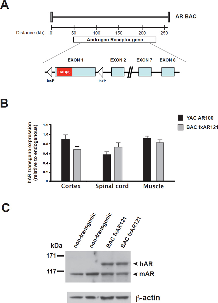
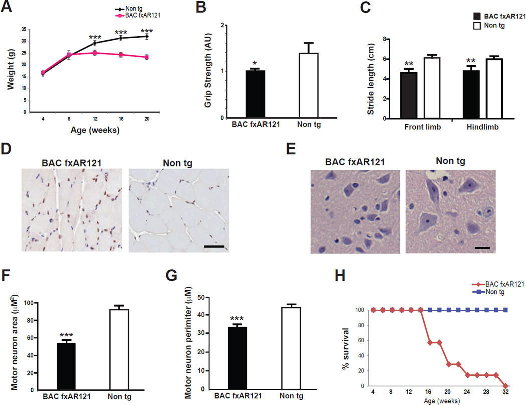
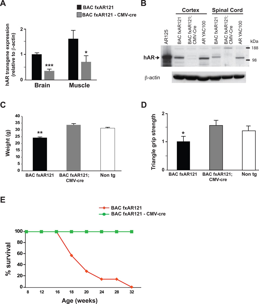
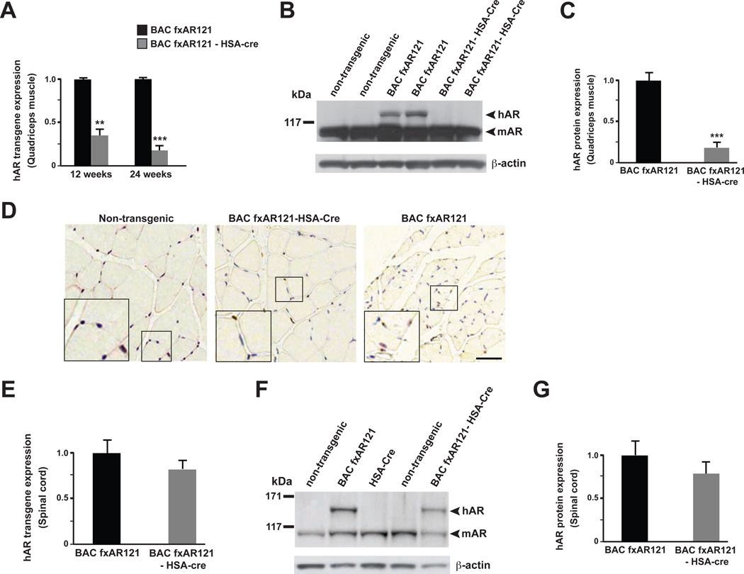
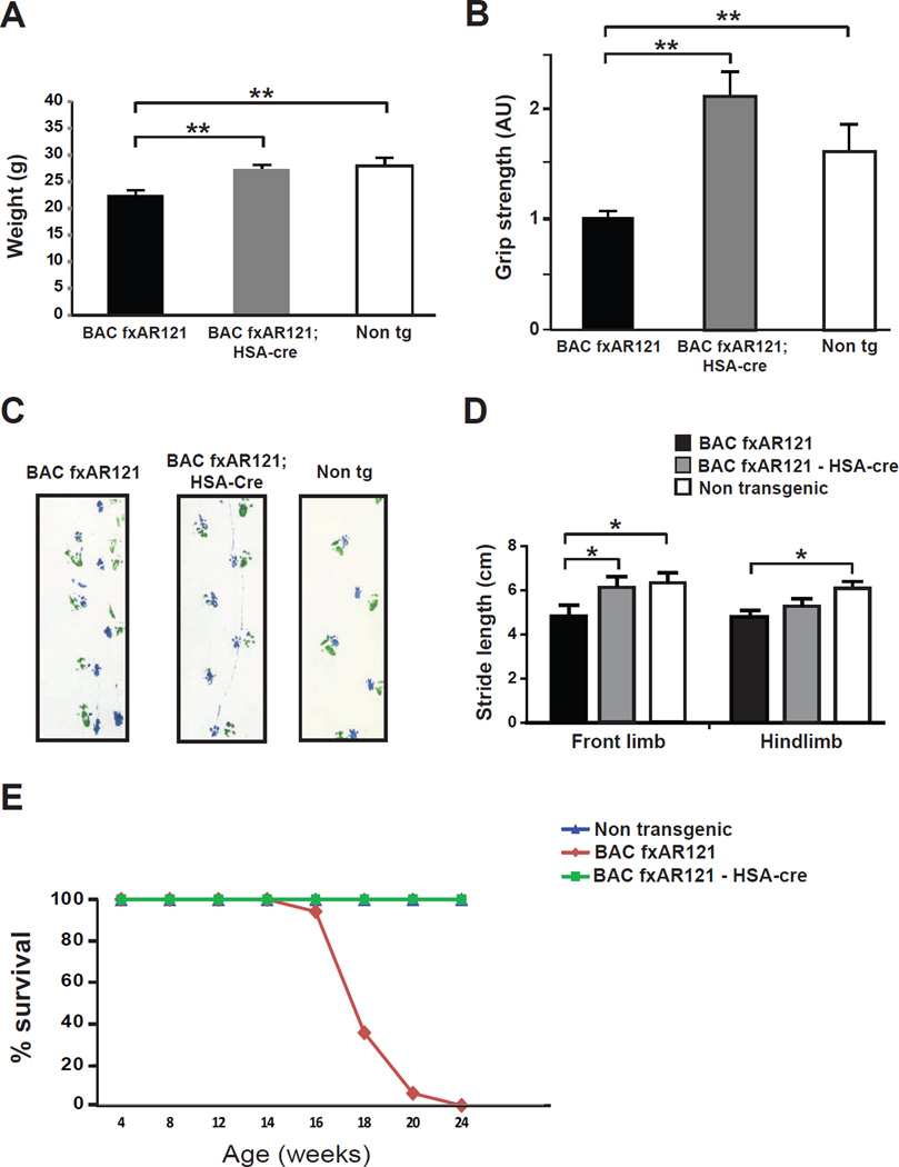
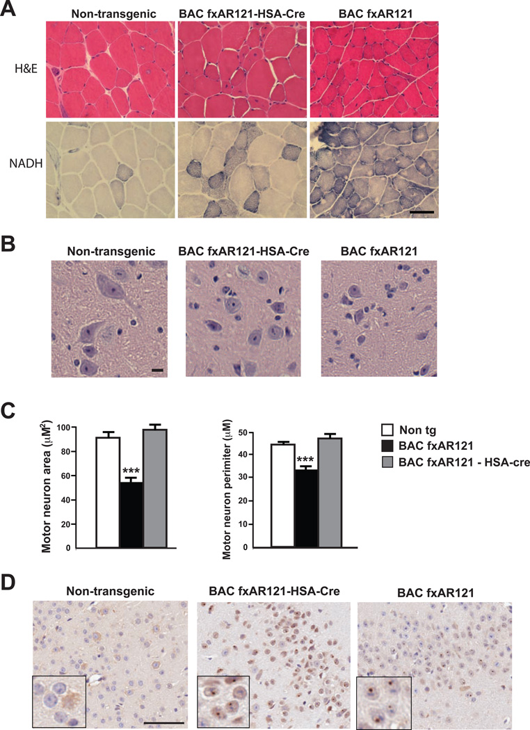
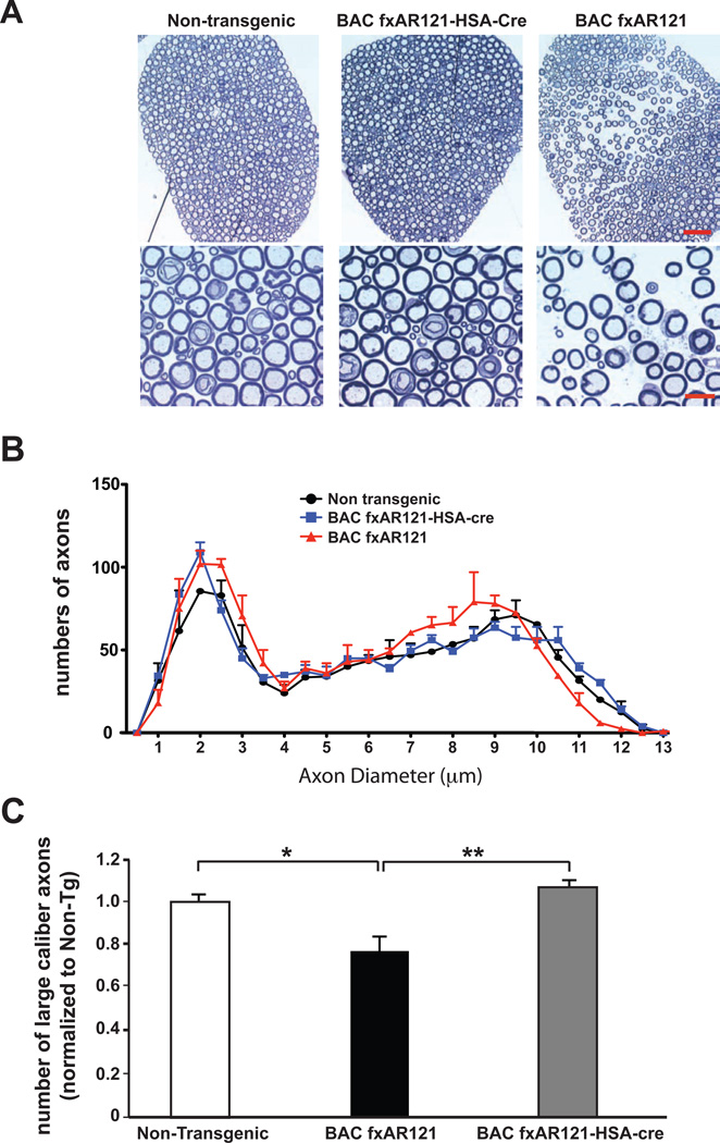
X-linked spinal and bulbar muscular atrophy (SBMA) is characterized by adult-onset muscle weakness and lower motor neuron degeneration. SBMA is caused by CAG-polyglutamine (polyQ) repeat expansions in the androgen receptor (AR) gene. Pathological findings include motor neuron loss, with polyQ-AR accumulation in intranuclear inclusions. SBMA patients exhibit myopathic features, suggesting a role for muscle in disease pathogenesis. To determine the contribution of muscle, we developed a BAC mouse model featuring a floxed first exon to permit cell-type-specific excision of human AR121Q. BAC fxAR121 mice develop systemic and neuromuscular phenotypes, including shortened survival. After validating termination of AR121 expression and full rescue with ubiquitous Cre, we crossed BAC fxAR121 mice with Human Skeletal Actin-Cre mice. Muscle-specific excision prevented weight loss, motor phenotypes, muscle pathology, and motor neuronopathy and dramatically extended survival. Our results reveal a crucial role for muscle expression of polyQ-AR in SBMA and suggest muscle-directed therapies as effective treatments.


Figures

Figure 1
Figure 1. Generation and expression analysis of BAC fxAR121 transgenic mice
(A) Diagram of BAC fxAR121 transgenic construct. After we generated a BAC containing all eight exons of the human Androgen Receptor (hAR) gene, we introduced a CAG121 repeat and two loxP sites flanking exon 1. (B) RT-PCR analysis of cortex, spinal cord, and quadriceps muscle hAR transgene RNA revealed expression levels that are slightly less than endogenous mouse AR RNA expression levels for BAC fxAR121 mice, and comparable to previously published YAC AR100 SBMA mice (Sopher et al., 2004). (C) Western blot analysis of whole brain protein lysates confirmed expression of full-length human AR protein (arrowhead, hAR) in the CNS of BAC fxAR121 mice. In this experiment, we used an anti-AR antibody that cross-reacts with mouse AR protein, which is detected at a lower molecular mass (arrowhead, mAR). β-actin immunoblotting serves as a loading control.
Figure 2
Figure 2. BAC fxAR121 transgenic mice display systemic and neuromuscular phenotypes
(A) By 12 weeks of age, male BAC fxAR121 mice no longer continue to gain weight, unlike their non-transgenic littermates (n = 6 – 14 / group). ***P < .001, t-test. (B) Combined grip strength analysis at 13 weeks of age reveals significant weakness in male BAC fxAR121 mice in comparison to non-transgenic littermate controls (n = 3 / group). Grip strength is given in arbitrary units, with BAC fxAR121 performance set to 1. *P < .05, t-test. (C) Measurement of front limb and hindlimb stride length was performed for 20 week-old male BAC fxAR121 mice and non-transgenic littermate controls (n = 5 – 6 / group). Significant decreases in mean stride length were observed for BAC fxAR121 mice in comparison to controls. **P < .01, t-test. (D) AR immunostaining of quadriceps muscle sections from 20 week-old male BAC fxAR121 mice and non-transgenic littermate controls reveals focal AR protein inclusions at the muscle cell periphery in BAC fxAR121 mice. These inclusions appear only occasionally in muscle sections from non-transgenic mice. Scale bar = 50 µm. (E) H&E-stained spinal cord sections from 28 week-old male BAC fxAR121 mice and non-transgenic littermate controls demonstrate that ventral horn neurons in the lumbar spinal cord are reduced in size in BAC fxAR121 males. Scale bar = 20 µm. (F) Quantification of ventral horn neuron soma size shown in (E), (n = 3 / group). ***P < .001, t-test. (G) Quantification of ventral horn neuron perimeter shown in (E), (n = 3 / group). ***P < .001, t-test. (H) Kaplan-Meier plot of male BAC fxAR121 mice and non-transgenic littermate controls (n = 9 / group) reveals a dramatic reduction in lifespan for BAC fxAR121 mice. P < .0001, Log-rank test. Error bars = s.e.m.
Figure 3
Figure 3. Ubiquitous Cre-mediated excision of the human AR121 transgene rescues systemic and neuromuscular phenotypes in BAC fxAR121 mice
(A) We measured hAR transgene expression in 6 week-old male BAC fxAR121 – CMV-Cre bigenic mice in comparison to singly transgenic male BAC fxAR121 mice (n = 3 / group) by qRT-PCR analysis. We observed marked reductions in hAR transgene expression in the brain and quadriceps muscle of BAC fxAR121 – CMV-Cre bigenic mice. Results are normalized to hAR transgene expression in BAC fxAR121 brain, which was set to 1. ***P < .001, *P < .05, t-test. (B) Western blot analysis of cortex and spinal cord for 6 week-old male BAC fxAR121 – CMV-Cre bigenic mice and littermate male BAC fxAR121 mice indicates that hAR protein expression is no longer detectable in BAC fxAR121 – CMV-Cre bigenic mice, when immunoblotted with human-specific anti-AR antibody. The first lane contains protein isolated from HEK293 cells transfected with a hAR125Q expression construct, and YAC AR100 protein lysates are included as an additional positive control. β-actin immunoblotting serves as a loading control. (C) Ubiquitous Cre expression in BAC fxAR121 mice rescues weight loss in 16 week-old male BAC fxAR121 – CMV-Cre bigenic mice (n = 4 – 10 / group). **P < .01, ANOVA with post-hoc Tukey test. (D) Ubiquitous Cre expression in BAC fxAR121 mice rescues grip strength in 13 week-old BAC fxAR121 – CMV-Cre bigenic mice (n = 4 / group). Grip strength is given in arbitrary units, with BAC fxAR121 performance set to 1. *P < .05, ANOVA with post-hoc Tukey test. (E) Kaplan-Meier plot of male BAC fxAR121 mice and male BAC fxAR121 – CMV-Cre bigenic mice (n = 9 / group) reveals significantly extended lifespan in BAC fxAR121 – CMV-Cre mice. P < .0001, Log-rank test. BAC fxAR121 – CMV-Cre bigenic mice are still alive and well beyond 24 months of age. Error bars = s.e.m.
Figure 4
Figure 4. Muscle-restricted inactivation of human AR transgene expression in BAC fxAR121 – HSA-Cre mice
(A) We measured hAR transgene expression in quadriceps muscle in male BAC fxAR121 – HSA-Cre bigenic mice in comparison to singly transgenic male BAC fxAR121 mice (n = 3 / group) by qRT-PCR analysis. We documented a marked reduction in hAR transgene expression in BAC fxAR121 – HSA-Cre mice at 12 weeks and 24 weeks of age. Transgene expression is given in arbitrary units, with BAC fxAR121 set to 1. **P < .01, ***P < .001, t-test. (B) Western blot analysis of quadriceps muscle for 12 week-old male BAC fxAR121 – HSA-Cre bigenic mice and littermate male BAC fxAR121 mice indicates that hAR protein expression (arrowhead) is extremely reduced in muscle from BAC fxAR121 – HSA-Cre mice, when immunoblotted with anti-AR antibody, which cross-reacts with mAR protein (arrowhead). β-actin immunoblotting serves as a loading control. (C) Densitometry quantification of muscle protein immunoblot data shown in (B), n = 4 separate experiments. ***P < .001, t-test. (D) AR immunostaining of quadriceps muscle sections from 28 week-old male BAC fxAR121 mice, BAC fxAR121 – HSA-Cre mice, and non-transgenic littermate controls reveals a marked reduction in AR protein inclusions at the muscle cell periphery in BAC fxAR121 – HSA-Cre mice. Scale bar = 50 µm. (E) We measured hAR transgene expression in spinal cord from 12 week-old male BAC fxAR121 – HSA-Cre bigenic mice and BAC fxAR121 mice (n = 3 / group) by qRT-PCR analysis, and observed similar hAR expression levels. P = n.s., t-test. Transgene expression is given in arbitrary units, with BAC fxAR121 set to 1. (F) Western blot analysis of spinal cord for 12 week-old male BAC fxAR121 – HSA-Cre bigenic mice and littermate male BAC fxAR121 mice indicates that hAR protein expression (arrowhead) is not reduced, relative to mouse AR protein expression (arrowhead) in BAC fxAR121 – HSA-Cre mice, when immunoblotted with anti-AR antibody, which cross-reacts with mouse AR protein. β-actin immunoblotting serves as a loading control. (G) Densitometry quantification of spinal cord protein immunoblot data shown in (F), n = 4 separate experiments. P = n.s., t-test. Error bars = s.e.m.
Figure 5
Figure 5. Cre-mediated excision of the human AR121 transgene from skeletal muscle rescues systemic and neuromuscular phenotypes in BAC fxAR121 mice
(A) Cre expression in skeletal muscle rescues weight loss in 20 week-old male BAC fxAR121 mice (n = 5 – 10 / group). **P < .01, ANOVA with post-hoc Tukey test. (B) Cre expression in skeletal muscle rescues grip strength in 20 week-old male BAC fxAR121 mice (n = 5 – 10 / group). Grip strength is given in arbitrary units, with BAC fxAR121 performance set to 1. **P < .01, ANOVA with post-hoc Tukey test. (C) Footprint analysis of 20 week-old male BAC fxAR121 mice, BAC fxAR121 – HSA-Cre mice, and littermate non-transgenic controls reveal normal stride in non-transgenic individuals (right), but obviously shortened strides in BAC fxAR121 mice (left). The stride of BAC fxAR121 – HSA-Cre mice is visibly improved (middle), more closely resembling the performance of non-transgenic mice. Fore paw prints are green, and hind paw prints are blue. (D) Quantification of stride length analysis shown in (C), (n = 6 – 8 / group). *P < .05, ANOVA with post-hoc Tukey test, front limb. *P < .05, ANOVA with post-hoc Tukey test, hindlimb: Non tg vs. BAC fxAR121, P < .05. BAC fxAR121-HSA-Cre vs. BAC fxAR121, P = n.s. (E) Kaplan-Meier plot of male BAC fxAR121 mice, male BAC fxAR121 – HSA-Cre bigenic mice, and male non-transgenic littermates (n = 9 – 13 / group) reveals significantly extended lifespan in BAC fxAR121 – HSA-Cre mice. P < .0001, Log-rank test. BAC fxAR121 – HSA-Cre bigenic mice are still alive and well beyond 16 months of age. Error bars = s.e.m.
Figure 6
Figure 6. Excision of human AR transgene from skeletal muscle rescues neuromuscular pathology without reducing AR protein aggregate burden in CNS of BAC fxAR121 mice
(A) Quadriceps muscle sections from 18 week-old male non-transgenic littermate controls, BAC fxAR121 – HSA-Cre mice, and BAC fxAR121 mice were stained with hematoxylin and eosin (H&E) and NADH. H&E staining of non-transgenic muscle (left) shows normal fibers of similar shape and size, but BAC fxAR121 muscle (right) reveals significant atrophy, with most fibers of markedly reduced caliber. Sections of BAC fxAR121 – HSA-Cre muscle (middle) appear relatively normal, with most fibers of large caliber, though one area of smaller fibers is present. NADH staining of non-transgenic muscle (left) yields occasional dark-staining fibers, while NADH staining of BAC fxAR121 sections (right) uncovers collections of very dark-staining fibers, undergoing atrophy. Evidence for muscle fiber atrophy is only occasionally present in BAC fxAR121 – HSA-Cre mice (middle). Scale bar = 50 µm. (B) H&E-stained spinal cord sections from 28 week-old male BAC fxAR121 mice, BAC fxAR121 – HSA-Cre mice, and non-transgenic littermate controls reveals that ventral horn neurons in the lumbar spinal cord are reduced in size in BAC fxAR121 males, and that this size reduction phenotype is rescued in BAC fxAR121 – HSA-Cre mice. Scale bar = 50 µm. (C) Quantification of ventral horn neuron soma size and perimeter shown in (B), n = 3 / group. ***P < .001, ANOVA with post-hoc Tukey test. Error bars = s.e.m. (D) AR immunostaining of frontal cortex from 28 week-old male BAC fxAR121 mice, BAC fxAR121 – HSA-Cre mice, and non-transgenic littermates reveals prominent nuclear inclusions in the cortex of both BAC fxAR121 and BAC fxAR121 – HSA-Cre mice (see inset for AR nuclear aggregates, which are visualized as dark brown puncta). Scale bar = 50 µm.
Figure 7
Figure 7. Excision of human AR transgene from skeletal muscle rescues motor neuron axon degeneration in BAC fxAR121 mice
(A) We sectioned L5 spinal cord root motor axons, and observed increased spacing in 28 week-old male BAC fxAR121 mice (right). No evidence of axon spacing was present in micrographs from non-transgenic mice (left) or BAC fxAR121 – HSA-Cre littermates (middle). Scale bar, top panel = 50 µm. Scale bar, bottom = 10 µm. (B) Mean diameters of L5 spinal cord root motor axons in 28 week-old male non-transgenic mice, BAC fxAR121 – HSA-Cre mice, and BAC fxAR121 mice. (C) Counting of motor axons revealed a significant reduction in large caliber (> 9 µm diameter) axons in BAC fxAR121 mice. This phenotype is rescued in BAC fxAR121 – HSA-Cre bigenic mice. **P < .01, ANOVA with post-hoc Tukey test: P < .01 for BAC fxAR121-HSA-Cre vs. BAC fxAR121, P < .05 for Non-transgenic vs. BAC fxAR121. Error bars = s.e.m.

Comment in

  • Muscle matters in Kennedy's disease.
    Rinaldi C, Bott LC, Fischbeck KH. Rinaldi C, et al. Neuron. 2014 Apr 16;82(2):251-3. doi: 10.1016/j.neuron.2014.04.005. Neuron. 2014. PMID: 24742452 Free PMC article.

References

    1. Arnold ES, Ling SC, Huelga SC, Lagier-Tourenne C, Polymenidou M, Ditsworth D, Kordasiewicz HB, McAlonis-Downes M, Platoshyn O, Parone PA, et al. ALS-linked TDP-43 mutations produce aberrant RNA splicing and adult-onset motor neuron disease without aggregation or loss of nuclear TDP-43. Proc Natl Acad Sci U S A. 2013;110:E736–E745. - PMC - PubMed
    1. Azzouz M, Ralph GS, Storkebaum E, Walmsley LE, Mitrophanous KA, Kingsman SM, Carmeliet P, Mazarakis ND. VEGF delivery with retrogradely transported lentivector prolongs survival in a mouse ALS model. Nature. 2004;429:413–417. - PubMed
    1. Benatar M, Wuu J, Fernandez C, Weihl CC, Katzen H, Steele J, Oskarsson B, Taylor JP. Motor neuron involvement in multisystem proteinopathy: Implications for ALS. Neurology. 2013;80:1874–1880. - PMC - PubMed
    1. Boillee S, Yamanaka K, Lobsiger CS, Copeland NG, Jenkins NA, Kassiotis G, Kollias G, Cleveland DW. Onset and progression in inherited ALS determined by motor neurons and microglia. Science. 2006;312:1389–1392. - PubMed
    1. Bosch-Marce M, Wee CD, Martinez TL, Lipkes CE, Choe DW, Kong L, Van Meerbeke JP, Musaro A, Sumner CJ. Increased IGF-1 in muscle modulates the phenotype of severe SMA mice. Hum Mol Genet. 2011;20:1844–1853. - PMC - PubMed

Publication types

MeSH terms