. 2014 Jul;190(1):55-63.
doi: 10.1016/j.jss.2014.03.036. Epub 2014 Mar 20.

Oridonin inhibits hepatic stellate cell proliferation and fibrogenesis

Affiliations

Oridonin inhibits hepatic stellate cell proliferation and fibrogenesis

Fredrick J Bohanon et al. J Surg Res. 2014 Jul.

Abstract

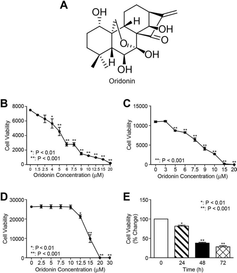
Background: Liver fibrosis is a common response to liver injury and, in severe cases, leads to cirrhosis. The hepatic stellate cells (HSCs) become activated after liver injury and play a significant role in fibrogenesis. The activated HSC is characterized by increased proliferation, overexpression of α smooth muscle actin, and excessive production of extracellular matrix (ECM) proteins. Oridonin, a naturally occurring diterpenoid, has been shown to induce apoptosis in liver and gastric cancer cells. However, its effects on the HSC are unknown.

Methods: We tested the effects of oridonin on the activated human and rat HSC lines LX-2 and HSC-T6, and the human hepatocyte cell line C3A. Transforming growth factor β1 (TGF-β1) was used to stimulate LX-2 cells.

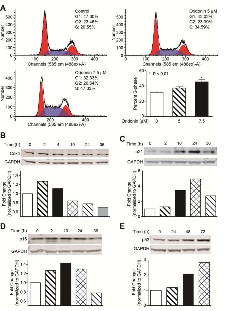
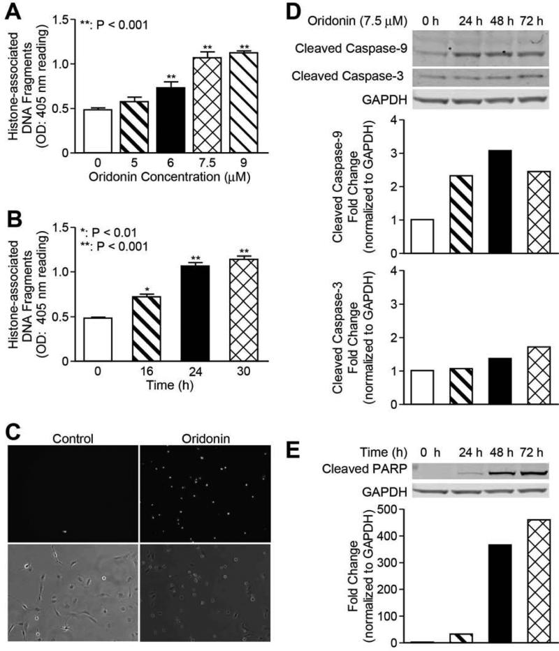
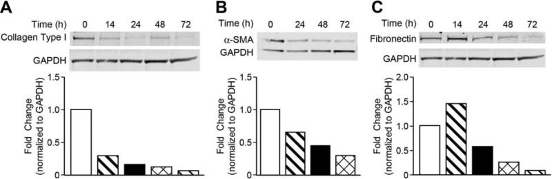
Results: Oridonin significantly inhibited LX-2 and HSC-T6 proliferation. In contrast, oridonin had no antiproliferative effect on C3A cells at our tested range. Oridonin induced apoptosis and S-phase arrest in LX-2 cells. These findings were associated with an increase in p53, p21, p16, and cleaved Poly (ADP-ribose) Polymerase (PARP), and with a decrease in Cyclin-dependent kinase 4 (Cdk4). Oridonin markedly decreased expression of α smooth muscle actin and ECM protein type I collagen and fibronectin, blocked TGF-β1-induced Smad2/3 phosphorylation and type I collagen expression.

Conclusions: Oridonin induces apoptosis and cell cycle arrest involving the p53-p21 pathway in HSC and appears to be nontoxic to hepatocytes. In addition, oridonin suppressed endogenous and TGF-β1-induced ECM proteins. Thus, oridonin may act as a novel agent to prevent hepatic fibrosis.

Keywords: Apoptosis; Liver fibrosis; Oridonin; Stellate cells.


Figures

Fig. 1
Fig. 1. Oridonin suppresses HSC proliferation
Chemical structure of oridonin (A). LX-2 cells (B), HSC-T6 cells (C) and C3A hepatocytes (D) were treated with a series of concentrations of oridonin for 48 hours, and cell viability was determined using Alamar Blue assay. (E) LX-2 cells were treated with 7.5 μmol/L of oridonin for 24, 48, and 72 hours; cell viability was measured by Alamar Blue assay. P-values shown compared to vehicle (0.1% DMSO, 0 μmol/L). The results are representative of at least three independent experiments.
Fig. 2
Fig. 2. Oridonin induces LX-2 cell cycle arrest
(A) Oridonin induces S-phase cell cycle arrest. LX-2 cells were treated with oridonin (0, 5, and 7.5 μmol/L) for 24 hours. Cells were stained with propidium iodide and analyzed by flow cytometry as described in Methods. The experiments were repeated three times and representative data are shown. (B), (C), (D), and (E) Oridonin affects cell cycle regulatory proteins. LX-2 cells were treated with vehicle (0.1% DMSO) or oridonin (7.5 μmol/L) for indicated time points. Whole cell lysates were analyzed by Western blot with antibodies for Cdk4, p21, p16, and p53. GAPDH was used as loading control. Densitometric analyses of bands were quantified and data expressed as fold change of control normalized to GAPDH.
Fig. 3
Fig. 3. Oridonin promotes LX-2 cell apoptosis
LX-2 cells were incubated with different concentrations or time points as indicated. Apoptosis by oridonin was evaluated either by Cell Death detection ELISA (each conducted in triplicate) (A) and (B), or Yo-Pro-1 staining (C). Whole cell lysates were analyzed by Western blot with antibodies for cleaved caspase-3 and cleaved caspase-9 (D), cleaved-PARP (E). GAPDH was used as loading control. Densitometric analyses of bands were quantified and data expressed as fold of control normalized to GAPDH. The results are representative of at least three independent experiments.
Fig. 4
Fig. 4. Oridonin suppresses α-SMA and ECM protein expression
LX-2 cells were incubated with oridonin (7.5 μmol/L) at time points as indicated. Whole cell lysates were analyzed by Western blot with antibodies for Type I collagen (A), α-Smooth muscle actin (B) and fibronectin (C). GAPDH was used as loading control. Densitometric analyses of bands were quantified and data expressed as fold change of control normalized to GAPDH. The results are representative of at least three independent experiments.
Fig. 5
Fig. 5. Oridonin inhibits TGF-β induced pSmad activity and ECM
LX-2 cells were preincubated with oridonin (7.5 μmol/L) for 2 hours and then treated with TGF-β (2 ng/mL) for 18 hours, whole cell lysates were analyzed by Western blot with antibodies for phosphorylated-Smad2/3 (A) and type I collagen (B), fibronectin (C). GAPDH was used as loading control. Densitometric analyses of bands were quantified and data expressed as fold change of control normalized to GAPDH. (D) LX-2 cells were pretreated with 7.5 mol/L of oridonin for 1 hour and then treated with TGF-β (2 ng/mL) for 1 hour. Nuclear proteins were extracted and analyzed by EMSA with 32P-labeled SBE probe described in experiment procedures.

References

    1. Neff GW, Duncan CW, Schiff ER. The current economic burden of cirrhosis. Gastroenterol Hepatol (N Y) 2011;7:661–671. - PMC - PubMed
    1. Lee UE, Friedman SL. Mechanisms of hepatic fibrogenesis. Best Pract Res Clin Gastroenterol. 2011;25:195–206. - PMC - PubMed
    1. Asahina K. Hepatic stellate cell progenitor cells. J Gastroenterol Hepatol. 2012;27(Suppl 2):80–84. - PMC - PubMed
    1. Lemoinne S, Cadoret A, El Mourabit H, Thabut D, Housset C. Origins and functions of liver myofibroblasts. Biochim Biophys Acta. 2013;1832:948–954. - PubMed
    1. Tsukamoto H, Zhu NL, Wang J, Asahina K, Machida K. Morphogens and hepatic stellate cell fate regulation in chronic liver disease. J Gastroenterol Hepatol. 2012;27(Suppl 2):94–98. - PMC - PubMed

Publication types

MeSH terms