

USP6 gene rearrangements occur preferentially in giant cell reparative granulomas of the hands and feet but not in gnathic location
- PMID: 24742829
- PMCID: PMC4225080
- DOI: 10.1016/j.humpath.2014.01.020
USP6 gene rearrangements occur preferentially in giant cell reparative granulomas of the hands and feet but not in gnathic location
Abstract
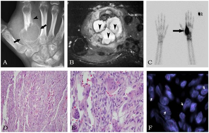
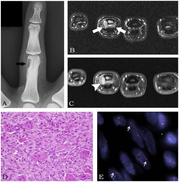
Giant cell reparative granulomas (GCRGs) are lytic lesions that occur predominantly in the gnathic bones and occasionally in the small bones of the hands and feet. They are morphologically indistinguishable from, and are regarded as synonymous with, solid variant of aneurysmal bone cysts (ABC) in extragnathic sites. Identification of USP6 gene rearrangements in primary ABC has made possible investigating potential pathogenetic relationships with other morphologic mimics. USP6 gene alterations in giant cell-rich lesions (GCRG/ABC) of small bones of the hands and feet have not been previously studied. We investigated USP6 gene alterations in a group of 9 giant cell-rich lesions of the hands and feet and compared the findings with morphologically similar lesions including 8 gnathic GCRGs, 22 primary ABCs, 8 giant cell tumors of bone, and 2 brown tumors of hyperparathyroidism. Overall, there were 49 samples from 48 patients including 26 females and 22 males. Of the 9 lesions of the hands and feet, 8 (89%) showed USP6 gene rearrangements, whereas no abnormalities were identified in the 8 gnathic GCRGs, 2 brown tumors, or 8 giant cell tumors of bone. Of the 22 primary ABCs, 13 (59%) showed USP6 gene rearrangements. In conclusion, most GCRGs of the hands and feet represent true ABCs and should be classified as such. The terminology of GCRG should be limited to lesions from gnathic location. Fluorescence in situ hybridization for USP6 break-apart is a useful ancillary tool in the diagnosis of primary ABCs and distinguishing them from GCRGs and other morphologically similar lesions.
Keywords: Giant cell reparative granuloma; Solid ABC; USP6.
Copyright © 2014 Elsevier Inc. All rights reserved.
Figures
References
-
- Jaffe HL. Giant-cell reparative granuloma, traumatic bone cyst, and fibrous (fibro-oseous) dysplasia of the jawbones. Oral surgery, oral medicine, and oral pathology. 1953;6:159–175. - PubMed
-
- Ackerman LV, Spjut HJ. Armed Forces Institute of Pathology. Washington D.C.: 1962. Tumors of bone and cartilage.
-
- Lorenzo JC, Dorfman HD. Giant-cell reparative granuloma of short tubular bones of the hands and feet. The American journal of surgical pathology. 1980;4:551–563. - PubMed
-
- Sanerkin NG, Mott MG, Roylance J. An unusual intraosseous lesion with fibroblastic, osteoclastic, osteoblastic, aneurysmal and fibromyxoid elements. "Solid" variant of aneurysmal bone cyst. Cancer. 1983;51:2278–2286. - PubMed
-
- Ratner V, Dorfman HD. Giant-cell reparative granuloma of the hand and foot bones. Clinical orthopaedics and related research. 1990:251–258. - PubMed
MeSH terms
Substances
Grants and funding
LinkOut - more resources
Full Text Sources
Other Literature Sources
