. 2014 Apr 30;5(8):2131-48.
doi: 10.18632/oncotarget.1852.

Inhibition of protein kinase II (CK2) prevents induced signal transducer and activator of transcription (STAT) 1/3 and constitutive STAT3 activation

Affiliations

Inhibition of protein kinase II (CK2) prevents induced signal transducer and activator of transcription (STAT) 1/3 and constitutive STAT3 activation

Samadhi Aparicio-Siegmund et al. Oncotarget. .

Abstract

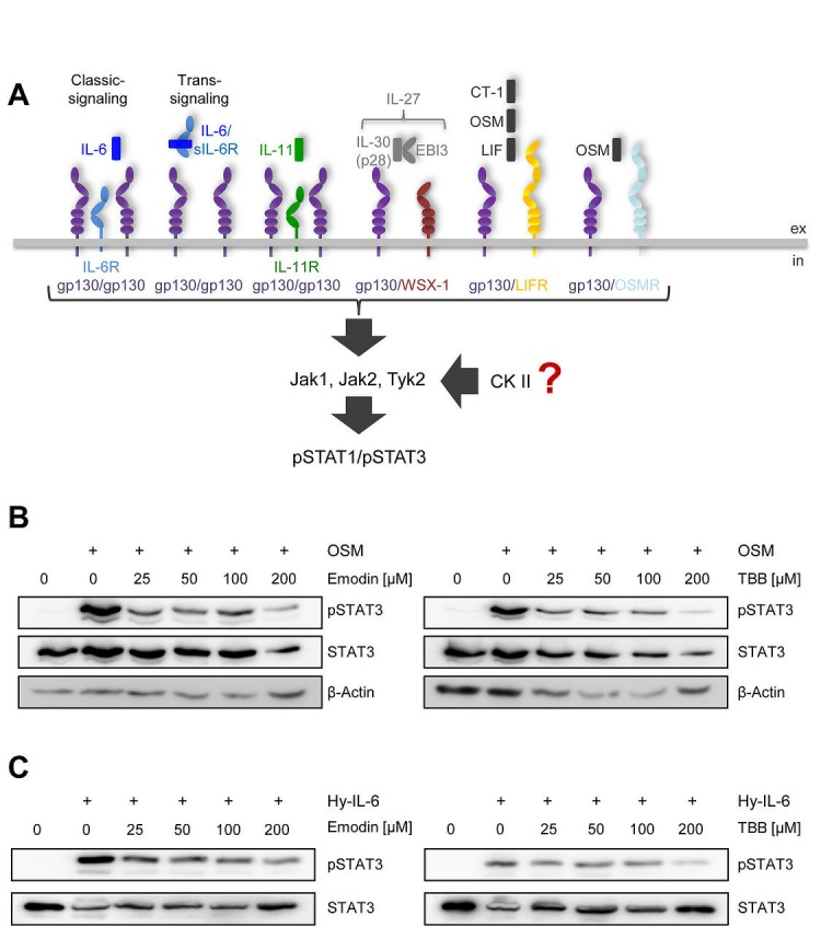
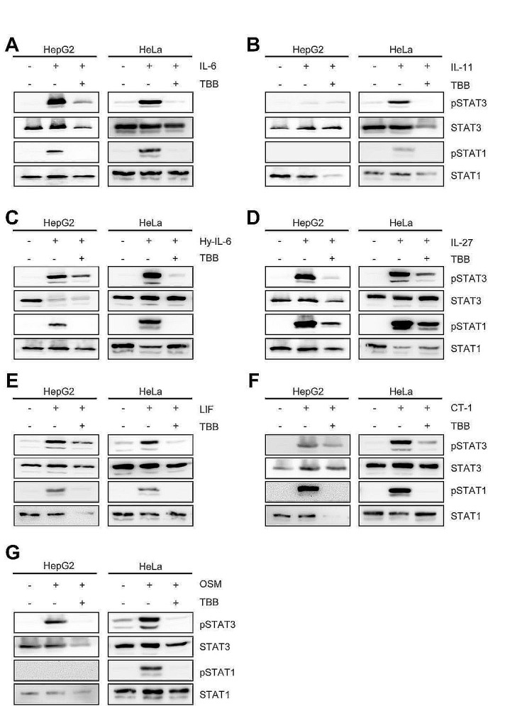
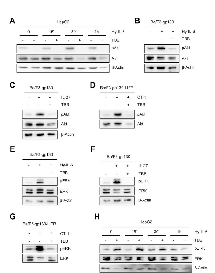
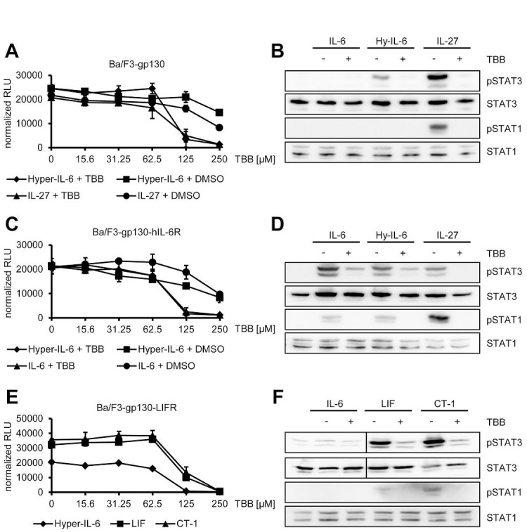
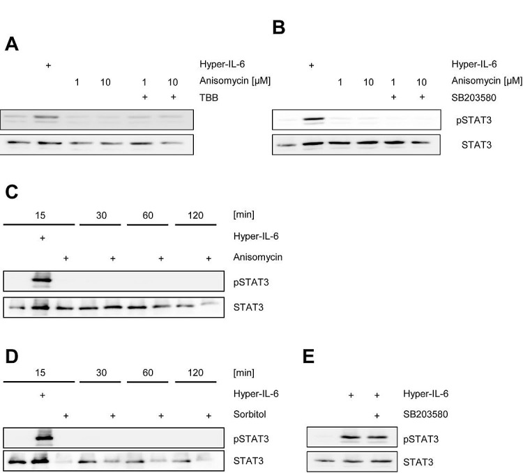
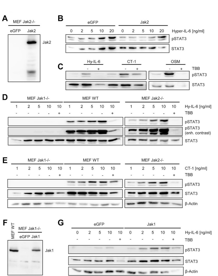
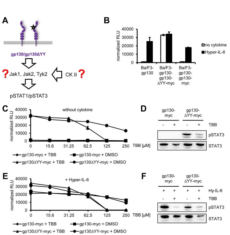
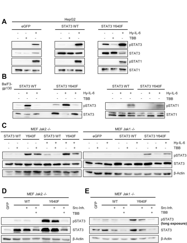
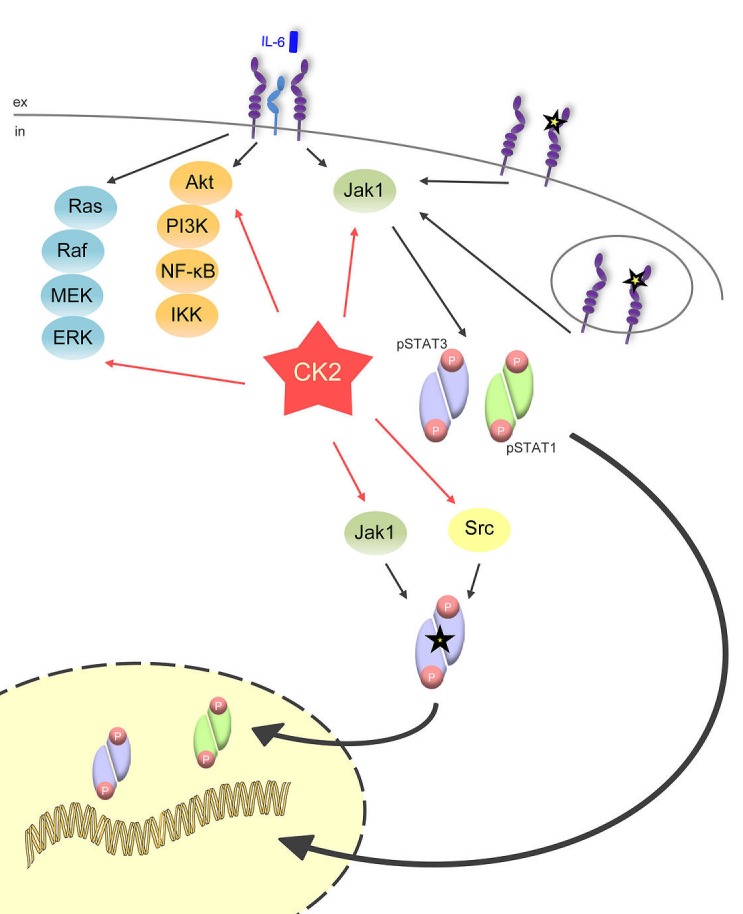
The Janus kinase / signal transducer and activator of transcription (Jak/STAT) pathway can be activated by many different cytokines, among them all members of the Interleukin (IL-)6 family. Dysregulation of this pathway, resulting in its constitutive activation, is associated with chronic inflammation and cancer development. In the present study, we show that activity of protein kinase II (CK2), a ubiquitously expressed serine/threonine kinase, is needed for induced activation of STAT1 and STAT3 by IL-6 classic and trans-signaling, IL-11, IL-27, oncostatin M (OSM), leukemia inhibitory factor (LIF) and cardiotrophin-1 (CT-1). Inhibition of CK2 efficiently prevented STAT phosphorylation and inhibited cytokine-dependent cell proliferation in a Jak1-dependent manner. Conversely, forced activation of CK2 alone was not sufficient to induce activation of the Jak/STAT signaling pathway. Inhibition of CK2 in turn inhibited Jak1-dependent STAT activation by oncogenic gp130 mutations. Furthermore, CK2 inhibition diminished the Jak1- and Src kinase-dependent phosphorylation of a constitutively active STAT3 mutant recently described in human large granular lymphocytic leukemia. In conclusion, we characterize CK2 as an essential component of the Jak/STAT pathway. Pharmacologic inhibition of this kinase is therefore a promising strategy to treat human inflammatory diseases and malignancies associated with constitutive activation of the Jak/STAT pathway.


Figures

Fig 1
Fig 1. CK2 is involved in STAT3 activation by OSM and Hyper-IL-6
(A) Schematic overview of the members of the IL-6 cytokine family and their receptors investigated in this study. IL-6 can activate a homodimer of glycoprotein 130 (gp130) either via the membrane-bound IL-6R (classic signaling) or via the soluble IL-6R (trans-signaling), whereas IL-11 acts only via a membrane-bound IL-11R. IL-27 (p28/IL-30 and EBI3) engages a gp130/WSX-1 heterodimer. The three members CT-1, OSM and LIF share a heterodimer of gp130/LIFR as signal transduction complex, while OSM can in addition also activate gp130 in combination with OSMR. IL-6 family cytokines activate the three kinases Jak1, Jak2 and Tyk2, which in turn phosphorylate STAT1 and STAT3. The influence of CK2 on this signaling pathway is investigated in the current study. (B) HepG2 cells were treated with different concentrations of the two CK2-inhibitors Emodin and TBB for 90 min. Cells were afterwards stimulated with 10 ng/ml OSM for 15 min. Phosphorylation of STAT3 was assessed by Western blotting. (C) HepG2 cells were treated as described under panel B, but were stimulated with 10 ng/ml Hyper-IL-6. Phosphorylation of STAT3 was assessed by Western blotting. One representative experiment of two performed is shown.
Fig 2
Fig 2. Inhibition of CK2 blocks IL-6 family induced STAT signaling
Serum-starved HepG2 or HeLa cells were stimulated with the indicated cytokines for 15 min: (A) HepG2: 10 ng/ml IL-6; HeLa: 20 ng/ml IL-6, (B) HepG2: 10 ng/ml IL-11; HeLa: 20 ng/ml IL-11, (C) 10 ng/ml Hyper-IL-6, (D) 10 ng/ml IL-27, (E) 10 ng/ml LIF, (F) 10 ng/ml CT-1 and (G) 10 ng/ml OSM. The cells were pre-incubated with the CK2 inhibitor TBB (100 μM) for 90 min where indicated. Phosphorylation of STAT1 and STAT3 was assessed by Western blotting, and STAT1/STAT3 served as internal loading control, respectively. One representative Western blot from at least three independent experiments is shown.
Fig 3
Fig 3. Inhibition of CK2 blocks ERK and Akt signaling
(A) HepG2 cells were stimulated with 10 ng/ml Hyper-IL-6, and cells were harvested after 0, 15, 30 and 60min. Cells were either pre-treated with 100 μM TBB for 90 min or with DMSO as control. Phosphorylation and expression level of Akt were assessed by Western blotting, and β-Actin served as internal loading control. (B, C) Ba/F3-gp130 cells were stimulated with 10 ng/ml Hyper-IL-6 or 10 ng/ml IL-27. Cells were either pre-treated with 100 μM TBB for 90 min or with DMSO as control. Phosphorylation of Akt was assessed by Western blotting. (D) The experiment was performed as described under panel C, but Ba/F3-gp130-LIFR cells were stimulated with 10 ng/ml CT-1. (E-G) The experiments were performed as described under panel B-D. Phosphorylation and expression level of ERK were assessed by Western blotting, and β-actin served as internal loading control. (H) HepG2 cells were stimulated with 10 ng/ml Hyper-IL-6, and cells were harvested after 0, 15, 30 and 60 min. Cells were either pre-treated with 100 μM TBB for 90 min or with DMSO as control. Phosphorylation and expression levels of ERK were assessed by Western blotting, and β-actin served as internal loading control. One representative Western blot from at least three independent experiments is shown.
Fig 4
Fig 4. Inhibition of CK2 prevents cytokine-dependent proliferation of Ba/F3-gp130 cells
(A) Equal amounts of Ba/F3-gp130 cells were incubated with 10 ng/ml Hyper-IL-6 or 10 ng/ml IL-27 and either increasing amounts of TBB (0-250 μM) or the corresponding amount of DMSO as control. Cellular proliferation was determined as described in Material and Methods. (B) Equal amounts of Ba/F3-gp130 cells were serum-starved for 3 h and either left untreated or where stimulated with 10 ng/ml IL-6, 10 ng/ml Hyper-IL-6 or 10 ng/ml IL-27. Where indicated, cells were pretreated with 100 μM TBB for 90 min prior to cytokine stimulation. Phosphorylation of STAT1 and STAT3 was assessed by Western blotting, and STAT1/STAT3 served as internal loading control, respectively. (C) Equal amounts of Ba/F3-gp130-hIL-6R cells were incubated with 10 ng/ml Hyper-IL-6 or 10 ng/ml IL-6 and either increasing amounts of TBB (0-250 μM) or the corresponding amount of DMSO as control. Cellular proliferation was determined as described in Material and Methods. (D) Equal amounts of Ba/F3-gp130-hIL-6R cells were treated as described in panel (B). Phosphorylation of STAT1 and STAT3 was assessed by Western blotting, and STAT1/STAT3 served as internal loading control, respectively. (E) Equal amounts of Ba/F3-gp130-LIFR cells were incubated with 10 ng/ml Hyper-IL-6, 10 ng/ml LIF or 10 ng/ml CT-1 and increasing amounts of TBB (0-250 μM). Cellular proliferation was determined as described in Material and Methods. (F) Equal amount of Ba/F3-gp130-LIFR cells were treated as described in panel (B). Phosphorylation of STAT1 and STAT3 was assessed by Western blotting, and STAT1/STAT3 served as internal loading control, respectively. Proliferation assays as well as Western Blots show one representative experiment of three performed.
Fig 5
Fig 5. Activation of CK2 does not induce Jak/STAT signaling
(A) HepG2 cells were serum starved for 3 h and stimulated with either 10 ng/ml Hyper-IL6 for 15 min, anisomycin for 30 min or left unstimulated. The CK2 inhibitor TBB was added 60 min prior to stimulation where indicated. (B) HepG2 cells were serum-starved for 3 h and stimulated with either 10 ng/ml Hyper-IL6 for 15 min, anisomycin for 30 min or left unstimulated. The p38 MAPK inhibitor SB203580 was added 60 min prior to stimulation where indicated. (C) HepG2 cells were serum-starved for 3 h and either left untreated, were stimulated with 10 ng/ml Hyper-IL-6 or 10 μM anisomycin for 15 min. Furthermore, either untreated or anisomycin-stimulated cells were harvested after 30, 60 or 120 min. (D) HepG2 cells were serum-starved for 3 h and either left untreated, were stimulated with 10 ng/ml Hyper-IL-6 or 500 mM sorbitol for 15 min. Furthermore, either untreated or sorbitol-stimulated cells were harvested after 30, 60 or 120 min. (E) HepG2 cells were serum-starved for 3 h and stimulated with either 10 ng/ml Hyper-IL-6 for 15 min or left unstimulated. The p38 MAPK inhibitor SB203580 was added 60 min prior to stimulation where indicated. Phosphorylation of STAT1 and STAT3 was assessed by Western blotting, and STAT1/STAT3 served as internal loading control, respectively. Shown is one representative Western blot from at least three independent experiments.
Fig 6
Fig 6. Jak1, but not Jak2, is required for STAT activation by IL-6 family cytokines
(A) Jak2−/− MEFs were transiently transfected with a plasmid coding for eGFP or Jak2. Expression of Jak2 was assessed by Western blotting 48 h later. (B) Jak2−/− MEFs transiently transfected with eGFP or Jak2 were stimulated with increasing concentrations of Hyper-IL-6 (0-20 ng/ml) for 15 min. (C) Jak2−/− MEFs transiently transfected with Jak2 were stimulated with 10 ng/ml Hyper-IL-6, CT-1 or OSM for 15 min. Cells were pre-treated with 100 μM TBB for 90 min where indicated. (D, E) Jak1−/−, Jak2−/− and wildtype MEFs were stimulated with different concentrations (0-10 ng/ml) of Hyper-IL-6 or CT-1 for 15 min. Cells were pre-treated with 100 μM TBB for 90 min where indicated. (F) Jak1−/− MEFs were transiently transfected with a plasmid coding for eGFP or Jak1. Expression of Jak2 was assessed by Western blotting 48 h later. Lysate of wildtype MEFs served as positive control. (G) Jak1−/− MEFs transiently transfected with eGFP or Jak2 were stimulated with increasing concentrations of Hyper-IL-6 (0-20 ng/ml) for 15 min. Cells were pre-treated with 100 μM TBB for 90 min where indicated. Phosphorylation of STAT3 was determined in all experiments by Western blotting, and STAT3 as well as β-actin served as loading controls. Shown is one representative Western blot from at least three independent experiments.
Fig 7
Fig 7. Jak1-dependent STAT-activation of the constitutively active gp130ΔYY-mutant depends on CK2
(A) Schematic representation of the gp130ΔYY-mutant harboring a deletion in domain 2. The kinase leading to constitutive STAT activation and the role of CK2 in this are unknown. (B) Equal amounts of Ba/F3-gp130, Ba/F3-gp130-gp130-myc and Ba/F3-gp130-gp130ΔYY-myc cells were incubated with or without 10 ng/ml Hyper-IL-6. (C) Equal amounts of Ba/F3-gp130-gp130-myc and Ba/F3-gp130-gp130ΔYY-myc cells were incubated with either increasing amounts of TBB (0-250 μM) or the corresponding amount of DMSO as control without any cytokine. (D) Equal amounts of Ba/F3-gp130-gp130-myc and Ba/F3-gp130-gp130ΔYY-myc cells were serum-starved for 3 h, and cells were pretreated with 100 μM TBB for 90 min prior to cytokine stimulation where indicated. Phosphorylation of STAT1 and STAT3 was assessed by Western blotting, and STAT1/STAT3 served as internal loading control, respectively. (E) Cells were treated as described under panel (B), but 10 ng/ml Hyper-IL-6 was added. (F) Cells were treated as described under panel (D), but cells were stimulated with 10 ng/ml Hyper-IL-6 for 15min. Phosphorylation of STAT1 and STAT3 was assessed by Western blotting, and STAT1/STAT3 served as internal loading control, respectively. Cellular proliferation in all assays shown was determined as described in Material and Methods. Proliferation assays as well as Western Blots are one representative experiment of three performed.
Fig 8
Fig 8. Activity of CK2 is required for phosphorylation of a constitutively active STAT3 mutant
(A) HepG2 cells were transiently transfected with eGFP, STAT3wt or STAT3Y640F. Two days later, cells were serum-starved for 5h and stimulated with either 10 ng/ml Hyper-IL-6 or were pre-treated with 100 μM TBB for 90min where indicated. (B) Ba/F3-gp130 cells stably transduced with STAT3wt or STAT3Y640F were serum-starved for 3 h and stimulated with either 10 ng/ml Hyper-IL-6 or were pre-treated with 100 μM TBB for 90 min where indicated. Phosphorylation of STAT1 and STAT3 was determined by Western blotting, and STAT1/3 served as internal loading control. (C) Jak2−/− or Jak1−/− MEFs were transiently transfected with a plasmid coding for eGFP, STAT3wt or STAT3Y640F. Two days later, cells were serum-starved for 5 h and stimulated with either 10 ng/ml Hyper-IL-6 or were pre-treated with 75 μM TBB for 90 min where indicated. Phosphorylation of STAT3 was determined by Western blotting, and STAT3/β-actin served as internal loading control. (D, E) Jak2−/− or Jak1−/− MEFs were transiently transfected with a plasmid coding for eGFP, STAT3wt or STAT3Y640F. Two days later, cells were serum-starved for 5 h and treated with Src-inhibitor alone or Src-inhibitor in combination with TBB for 90 min. Phosphorylation of STAT3 was determined by Western blotting, and STAT3/β-actin served as internal loading control. Western Blots show one representative experiment of three performed.
Fig 9
Fig 9. CK2 is the central lynchpin of the signaling pathways investigated in this study
The schematic overview shows that IL-6 family cytokines activate the three major signaling pathways Jak/STAT, Ras/Raf/MEK/ERK, and Akt/PI3K. All of them need CK2 activity for the initiation of downstream signaling. The gp130ΔYY-mutant, which is constitutively active and signals from the cell membrane and from intracellular compartments, solely activates Jak/STAT signaling, and this can be efficiently blocked through CK2 blockade. A constitutively active STAT3 mutant (STAT3Y640F), which is constitutively phosphorylated by Jak1 and Src kinase, also needs CK2 activity

References

    1. Stark G, Darnell J. The JAK-STAT pathway at twenty. Immunity. 2012;36(4):503–514. - PMC - PubMed
    1. O'shea J, Plenge R. JAK and STAT Signaling Molecules in Immunoregulation and Immune-Mediated Disease. Immunity. 2012;36(4):542–550. - PMC - PubMed
    1. Garbers C, Hermanns H, Schaper F, Müller-Newen G, Grötzinger J, Rose-John S, Scheller J. Plasticity and cross-talk of Interleukin 6-type cytokines. Cytokine Growth Factor Rev. 2012;23(3):85–182. - PubMed
    1. Chen E, Staudt L, Green A. Janus kinase deregulation in leukemia and lymphoma. Immunity. 2012;36(4):529–541. - PMC - PubMed
    1. Casanova J-L, Holland S, Notarangelo L. Inborn errors of human JAKs and STATs. Immunity. 2012;36(4):515–528. - PMC - PubMed

Publication types