. 2014 Apr 17;9(4):e94106.
doi: 10.1371/journal.pone.0094106. eCollection 2014.

Soluble CEACAM8 interacts with CEACAM1 inhibiting TLR2-triggered immune responses

Affiliations

Soluble CEACAM8 interacts with CEACAM1 inhibiting TLR2-triggered immune responses

Bernhard B Singer et al. PLoS One. .

Abstract

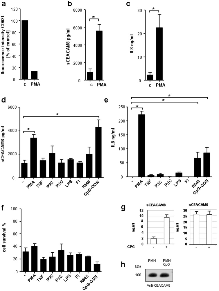
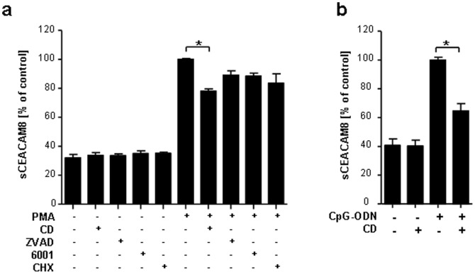
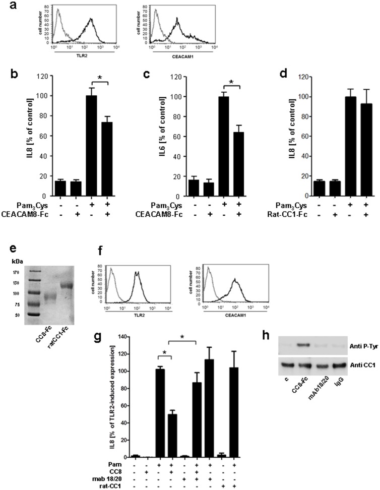
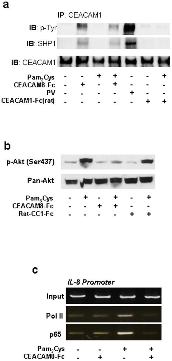
Lower respiratory tract bacterial infections are characterized by neutrophilic inflammation in the airways. The carcinoembryonic antigen-related cell adhesion molecule (CEACAM) 8 is expressed in and released by human granulocytes. Our study demonstrates that human granulocytes release CEACAM8 in response to bacterial DNA in a TLR9-dependent manner. Individuals with a high percentage of bronchial lavage fluid (BALF) granulocytes were more likely to have detectable levels of released CEACAM8 in the BALF than those with a normal granulocyte count. Soluble, recombinant CEACAM8-Fc binds to CEACAM1 expressed on human airway epithelium. Application of CEACAM8-Fc to CEACAM1-positive human pulmonary epithelial cells resulted in reduced TLR2-dependent inflammatory responses. These inhibitory effects were accompanied by tyrosine phosphorylation of the immunoreceptor tyrosine-based inhibitory motif (ITIM) of CEACAM1 and by recruitment of the phosphatase SHP-1, which could negatively regulate Toll-like receptor 2-dependent activation of the phosphatidylinositol 3-OH kinase-Akt kinase pathway. Our results suggest a new mechanism by which granulocytes reduce pro-inflammatory immune responses in human airways via secretion of CEACAM8 in neutrophil-driven bacterial infections.


Conflict of interest statement

Competing Interests: BBS has rights to a patent application related to CEACAMs. HS is a PLOS ONE Editorial Board member. There are no further patents, products in development or marketed products to declare. This does not alter the authors' adherence to all the PLOS ONE policies on sharing data and materials, as detailed online in the guide for authors. The patent name and numbers is the following: BBS: Use of soluble CEACAM8 for diagnosing, treating or monitoring diseases, and a method for screening compounds that prevent apoptosis. United States Patent 8,501,192 and European Patent Application No. 07117396.7-2123.

Figures

Figure 1
Figure 1. Analysis of CEACAM8 released by stimulated human granulocytes.
(a) In all experiments granulocytes were pre-incubated using 50 ng/ml GM-CSF for 90 min. Expression of CD62L on human granulocytes was assessed by FACS analysis after treatment with 20 ng/ml PMA for 1 h. Shown is fluorescence intensity as percent of control. Secreted CEACAM8 (sCEACAM8) (b) and IL8 (c) in supernatants of granulocytes incubated for 1 h with 20 ng/ml PMA measured by ELISA. (d) ELISA for released CEACAM8 in supernatants of granulocytes treated for 14 h with 20 ng/ml PMA, 5 ng/ml tumor necrosis factor α (TNFα), 10 µg/ml Pam3Cys (P3C), 100 ng/ml Poly(I∶C) (P∶IC), 1 µg/ml Flagellin (Fl), 10 µg/ml Resiquimod-848 (R848) or 100 µg/ml non-methylated CpG (CpG-ODN). (e) IL8 secretion measured by ELISA from supernatants of granulocytes treated the same way as in d. (f) Cells were stimulated with agonists like in (d) and (e) for 14 h. Cell viability (Annexin V/Propidium iodide) was determined by FACS analysis (f). Bars represent the mean of viable cells as assessed by Annexin V/Propidium jodide staining. (g) Soluble CEACAM6 (sCEACAM6) and CEACAM8 (sCEACAM8) in supernatants collected from 107 granulocytes treated for 14 hours with and without 100 µg/ml un-methylated CpG (CpG-ODN) were measured by ELISA. (h) To assess percentage of secreted CEACAM8 in relation to the total amount of cellular CEACAM8 lysates of CpG-ODN treated and untreated granulocyte were probed with the CEACAM8 specific mAb 80H3 by Western Blot. Data presented are mean ± s.e.m. of three different experiments performed in duplicates (b, c, d, e, f) or one of three identical experiments (a and h). * P<0.05.
Figure 2
Figure 2. Secretion of sCEACAM8 is inhibited by the actin inhibitor cytochalasin D.
(a) Released sCEACAM8 in supernatants of granulocytes measured by ELISA. Granulocytes were left untreated or pre-incubated for 1 h with one of the following stimuli: 0.5 µg/ml cytochalasin D (CD), 10 µM Pan caspase fmk Inhibitor Z-VAD (ZVAD), 10 µM Pan-MMP Inhibitor GM-6001 (6001) or 1 µg/ml cycloheximide (CHX) and then left untreated or incubated for 1 h with 20 ng/ml PMA. (b) CEACAM8 ELISA of supernatants harvested from granulocytes pre-treated with and without cytochalasin D (CD, 0.5 µg/ml) and 1 h stimulated with and without non-methylated CpG-ODN (b). Data presented are mean ± s.e.m. of three or four different experiments performed in duplicates. * P<0.05.
Figure 3
Figure 3. Soluble CEACAM8 interacts with membrane bound CEACAM1 and down-regulates the TLR2-triggered immune response on normal human bronchial epithelial (NHBE) cells.
(a) FACS analysis of CEACAM1 and TLR2 (black lines) expression on the cell surface of NHBE cells. Grey lines represent isotype-matched control mAb staining. (b, c and d) ELISA for IL8 and IL6, respectively, in supernatants of NHBE cells incubated with Pam3Cys alone, with Pam3Cys plus soluble CEACAM8-Fc and rat-CEACAM1-Fc, respectively (100 ng/ml each) or left untreated for 16 h. (e) 50 ng purified protein of CEACAM8-Fc and ratCEACAM1-Fc were analyzed by SDS-PAGE and stained by Coomassie blue to demonstrate equal amount and integrity of the used fusion proteins. (f) FACS analysis CEACAM1 and TLR2 expression (black lines) on the surface of A549 cells. Grey lines represent the isotype-matched control. (g) IL-8 released into the supernatants of A549 cells pre-incubated for 1 h with anti-CEACAM1 (mAb 18/20) or left untreated followed by stimulation with Pam3Cys alone or with Pam3Cys and CEACAM8-Fc and ratCEACAM1-Fc, respectively (100 ng/ml) for 16 h. Samples were measured by commercial ELISA. IL8 secretion following treatment of the cells with Pam3Cys served as positive control. (h) A549 cells were challenged either with CEACAM8-Fc or with mAb 18/20 (20 ng/ml). The IgG antibody was used as control. Cell lysates were assessed after CEACAM1 immunoprecipitation with anti-phospho tyrosine mAb 4G10 (upper panel). Then membrane was stripped and re-probed with anti-CEACAM1 mAb (lower panel) to measure the amount of precipitated CEACAM1. Data presented in (a, e, f and h) are from one experiment representative of three independent experiments, and the data presented in (b, c, d and g) are mean ± s.e.m. of three different experiments performed in triplicates, * P<0.05.
Figure 4
Figure 4. Interaction of soluble CEACAM8-Fc with CEACAM1 expressed on pulmonary epithelial cells mediates down-regulation of the TLR2-dependent immune response.
(a) FACS analysis of CEACAM1 (bold lines) expression on the cell surface of sparse A549-CEACAM1-4L and vector-transfected A549 cells (A549-vec). Thin lines represent isotype-matched control mAb stainings. (b) Sparsely grown A549-CEACAM1-4L cells were challenged with recombinant CEACAM8-Fc as indicated (white bars, left panel). Sparsely grown A549-vec cells were used as a control (black bars, left panel). Rat CEACAM1-Fc neither binds to sparse A549-CEACAM1-4L nor A549-vec cells. The presence and absence of CEACAM1 on the cell surface of sparse A549-vec and A549-CEACAM1-4L was tested utilizing CEACAM1 specific mAb B3-17 (right panel). Isotype matched IgG served as negative control. Black bars = A549-vec, white bars = A549-CEACAM1-4L. (c) Viability of A549 cells 16 h after treatment with CEACAM8-Fc, Pam3Cys (P3C), or with Pam3Cys plus soluble CEACAM8-Fc and ratCEACAM1-Fc, respectively (100 ng/ml each) or left untreated, were stained with annexin V-FITC/propidium iodide and measured using flow cytometry. (d) A549 cells were transfected with CEACAM1-targeting siRNA or control siRNA (csi) for 96 h and expression of CEACAM1 was measured by FACS analysis. (e) IL-8 released by A549 cells transfected for 96 h with CEACAM1-specific or control siRNA and infected for 16 h with Pam3Cys alone or in combination with CEACAM8-Fc (100 ng/ml) were measured in the supernatants of the cells by ELISA (e). (a, c and d) Data are from one experiment representative of three independent experiments. (b, and e) Data presented are mean ± s.e.m. of three different experiments performed in duplicates. *P<0.05.
Figure 5
Figure 5. The inhibitory effect of the soluble CEACAM8 interaction with membrane bound CEACAM1 for the TLR2-dependent immune response resembles the UspA1-CEACAM1 interaction of M. catarrhalis in pulmonary epithelial cells.
(a) Immunoprecipitation of CEACAM1 from A549 cell lysates. Cells were left untreated or were treated with Pam3Cys, Pam3Cys plus CEACAM8-Fc and ratCEACAM1-Fc, respectively (100 ng/ml). Stimulation with pervanadate served as positive control. Tyrosine phosphorylation of CEACAM1, co-precipitated SHP1 and total CEACAM1 were detected in this order by Western blot. (b) Immunoblot analysis of Akt phosphorylation in A549 cells treated with and without Pam3Cys alone or together with CEACAM8-Fc and ratCEACAM1-Fc, respectively (100 ng/ml each) for 60 min. Detection of Pan-Akt was used as loading control. (c) ChIP analysis of the binding of P65 and polymerase II (Pol II) to the IL8 promoter in A549 cells stimulated and unstimulated with Pam3Cys alone and together with CEACAM8-Fc (100 ng/ml). Data shown are from one experiment representative of three independent experiments.

Similar articles

Cited by

  • Fusobacterium nucleatum CbpF Mediates Inhibition of T Cell Function Through CEACAM1 Activation.
    Galaski J, Shhadeh A, Umaña A, Yoo CC, Arpinati L, Isaacson B, Berhani O, Singer BB, Slade DJ, Bachrach G, Mandelboim O. Galaski J, et al. Front Cell Infect Microbiol. 2021 Jul 15;11:692544. doi: 10.3389/fcimb.2021.692544. eCollection 2021. Front Cell Infect Microbiol. 2021. PMID: 34336716 Free PMC article.
  • The role of CEACAMs in neutrophil function.
    Skubitz KM. Skubitz KM. Eur J Clin Invest. 2024 Dec;54 Suppl 2(Suppl 2):e14349. doi: 10.1111/eci.14349. Eur J Clin Invest. 2024. PMID: 39674879 Free PMC article. Review.
  • Proteomic Bioprofiles and Mechanistic Pathways of Progression to Heart Failure.
    Ferreira JP, Verdonschot J, Collier T, Wang P, Pizard A, Bär C, Björkman J, Boccanelli A, Butler J, Clark A, Cleland JG, Delles C, Diez J, Girerd N, González A, Hazebroek M, Huby AC, Jukema W, Latini R, Leenders J, Levy D, Mebazaa A, Mischak H, Pinet F, Rossignol P, Sattar N, Sever P, Staessen JA, Thum T, Vodovar N, Zhang ZY, Heymans S, Zannad F. Ferreira JP, et al. Circ Heart Fail. 2019 May;12(5):e005897. doi: 10.1161/CIRCHEARTFAILURE.118.005897. Circ Heart Fail. 2019. PMID: 31104495 Free PMC article.
  • Moraxella catarrhalis induces CEACAM3-Syk-CARD9-dependent activation of human granulocytes.
    Heinrich A, Heyl KA, Klaile E, Müller MM, Klassert TE, Wiessner A, Fischer K, Schumann RR, Seifert U, Riesbeck K, Moter A, Singer BB, Bachmann S, Slevogt H. Heinrich A, et al. Cell Microbiol. 2016 Nov;18(11):1570-1582. doi: 10.1111/cmi.12597. Epub 2016 May 3. Cell Microbiol. 2016. PMID: 27038042 Free PMC article.
  • Immunoreceptors on neutrophils.
    van Rees DJ, Szilagyi K, Kuijpers TW, Matlung HL, van den Berg TK. van Rees DJ, et al. Semin Immunol. 2016 Apr;28(2):94-108. doi: 10.1016/j.smim.2016.02.004. Epub 2016 Mar 11. Semin Immunol. 2016. PMID: 26976825 Free PMC article. Review.

References

    1. Craig A, Mai J, Cai S, Jeyaseelan S (2009) Neutrophil recruitment to the lungs during bacterial pneumonia. Infect Immun 77: 568–575. - PMC - PubMed
    1. Parker D, Prince A (2011) Innate immunity in the respiratory epithelium. Am J Respir Cell Mol Biol 45: 189–201. - PMC - PubMed
    1. Mantovani A, Cassatella MA, Costantini C, Jaillon S (2011) Neutrophils in the activation and regulation of innate and adaptive immunity. Nat Rev Immunol 11: 519–531. - PubMed
    1. Schmidt T, Zundorf J, Gruger T, Brandenburg K, Reiners AL, et al. (2012) CD66b overexpression and homotypic aggregation of human peripheral blood neutrophils after activation by a gram-positive stimulus. J Leukoc Biol 91: 791–802. - PubMed
    1. Singer BB, Scheffrahn I, Heymann R, Sigmundsson K, Kammerer R, et al. (2002) Carcinoembryonic antigen-related cell adhesion molecule 1 expression and signaling in human, mouse, and rat leukocytes: evidence for replacement of the short cytoplasmic domain isoform by glycosylphosphatidylinositol-linked proteins in human leukocytes. J Immunol 168: 5139–5146. - PubMed

Publication types

MeSH terms