. 2014 Apr 17;9(4):e95365.
doi: 10.1371/journal.pone.0095365. eCollection 2014.

Uncoupling of glomerular IgA deposition and disease progression in alymphoplasia mice with IgA nephropathy

Affiliations

Uncoupling of glomerular IgA deposition and disease progression in alymphoplasia mice with IgA nephropathy

Masashi Aizawa et al. PLoS One. .

Abstract

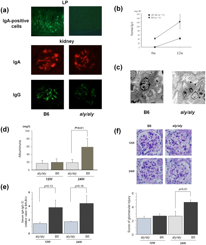
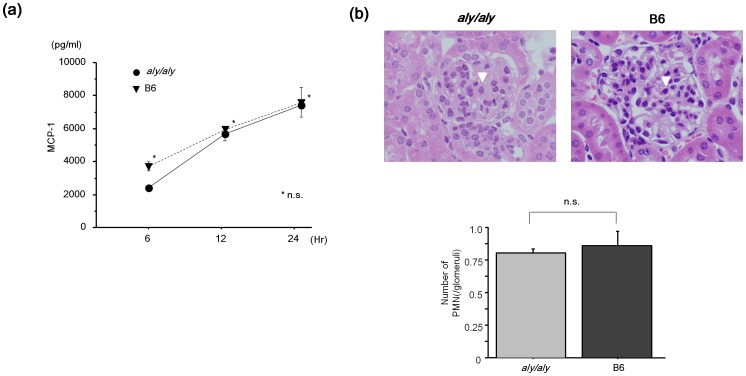
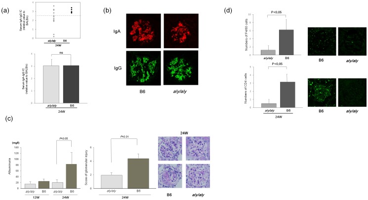
Previous clinical and experimental studies have indicated that cells responsible for IgA nephropathy (IgAN), at least in part, are localized in bone marrow (BM). Indeed, we have demonstrated that murine IgAN can be experimentally reconstituted by bone marrow transplantation (BMT) from IgAN prone mice in not only normal mice, but also in alymphoplasia mice (aly/aly) independent of IgA+ cells homing to mucosa or secondary lymphoid tissues. The objective of the present study was to further assess whether secondary lymph nodes (LN) contribute to the progression of this disease. BM cells from the several lines of IgAN prone mice were transplanted into aly/aly and wild-type mice (B6). Although the transplanted aly/aly showed the same degree of mesangial IgA and IgG deposition and the same serum elevation levels of IgA and IgA-IgG immune-complexes (IC) as B6, even in extent, the progression of glomerular injury was observed only in B6. This uncoupling in aly/aly was associated with a lack of CD4+ T cells and macrophage infiltration, although phlogogenic capacity to nephritogenic IC of renal resident cells was identical between both recipients. It is suggested that secondary LN may be required for the full progression of IgAN after nephritogenic IgA and IgA/IgG IC deposition.


Conflict of interest statement

Competing Interests: The authors have declared that no competing interests exist.

Figures

Figure 1
Figure 1. Reconstitution of IgA nephropathy by BM transplantation from ddY mice in aly/aly and B6 mice.
BM transplantation (BMT) from IgAN onset ddY mice (line A) (N = 24) reconstituted glomerular IgA (a) with serum elevation of IgA (b) in aly/aly and WT control B6 mice (aly/aly and B6 at 12 and 24 w, n = 6, each) independently of homing of IgA-positive cells into lamina propria (LP). Clear glomerular IgG deposition was found in B6, but not in aly/aly mice. Although electron-dense deposits (arrow head) were detected in aly/aly mice at 24 w after BMT as well as B6 mice (c), progression of urinary protein (d) and glomerular lesions (f) were absent in aly/aly mice in association with the lack of serum elevation of IgA-IgG immune complex (IC) (e). Data are presented as mean ± SD.
Figure 2
Figure 2. Identical phlogogenic capacity of renal resident cells in aly/aly and B6 mice.
(a) To confirm the nephritogenic capacity to of renal resident cells in aly/aly and B6 mice, chemokine expressions, such as MCP-1, were examined with mesangial cells from both mice under stimulation with IgA IC. MCP-1 concentrations in supernatants of both mice were not significantly different (n = 3, each). (b) Next to confirm phlogonenic capacity of aly/aly mice, anti-GBM glomerulonephritis was induced in aly/aly and B6 mice (n = 3 each). An influx of PMN (arrow head) was shown in the glomerulus of both mice. The number of PMN in the glomerulus of aly/aly and B6 mice were not significantly different. Data are presented as mean ± SD.
Figure 3
Figure 3. Uncoupling of glomerular IgA and disease progression in transplanted aly/aly mice was independent of IgA-IgG IC formation and deposition.
To further assess underlying mechanisms in the lack of the disease progression of IgAN in aly/aly mice, BM cells from a different line of poor-prognosis IgAN prone (line B) were transplanted in both mice. Although the serum levels of IgA-IgG IC were variable, some of the transplanted aly/aly mice (n = 5) showed similar amounts of IC (>2.5) to B6 mice (n = 5) (a). These selected aly/aly mice showed clear glomerular deposition of not only IgA but also IgG at 24 w, as seen in B6 mice (b). However, the progression of proteinuria and glomerular lesions was absent (c). This was associated with lack of F4/80 positive macrophages and CD4+ T cells (d). Data are presented as mean ± SD.

References

    1. Burger J, Hinglais N (1968) Les Depots intercapillaries d’IgA-IgG. Urol Nephrol Paris 74: 694–695. - PubMed
    1. Tomino Y, Endoh M, Nomoto Y, Sakai H (1981) Immunoglobulin A1 and IgA nephropathy. N Engl J Med. 305: 1159–1160. - PubMed
    1. Barratt J, Feehally J (2005) IgA nephropathy. J Am Soc Nephrol 16: 2088–2097. - PubMed
    1. van den Wall Bake AW, Daha MR, Evers-Schouten J, van Es LA (1988) Serum IgA and the production of IgA by peripheral blood and bone marrow lymphocytes in patients with primary IgA nephropathy: evidence for the bone marrow as the source of mesangial IgA. Am J Kidney Dis 12: 410–414. - PubMed
    1. Harper SJ, Allen AC, Pringle JH, Feehally J (1996) Increased dimeric IgA producing B cells in the bone marrow in IgA nephropathy determined by in situ hybridisation for J chain mRNA. J Clin Pathol 49: 38–42. - PMC - PubMed

Publication types