. 2014 Apr 17;9(4):e95588.
doi: 10.1371/journal.pone.0095588. eCollection 2014.

Curcumin significantly enhances dual PI3K/Akt and mTOR inhibitor NVP-BEZ235-induced apoptosis in human renal carcinoma Caki cells through down-regulation of p53-dependent Bcl-2 expression and inhibition of Mcl-1 protein stability

Affiliations

Curcumin significantly enhances dual PI3K/Akt and mTOR inhibitor NVP-BEZ235-induced apoptosis in human renal carcinoma Caki cells through down-regulation of p53-dependent Bcl-2 expression and inhibition of Mcl-1 protein stability

Bo Ram Seo et al. PLoS One. .

Erratum in

Abstract

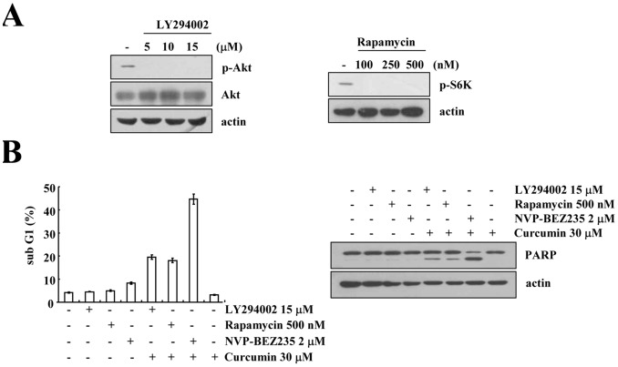
The PI3K/Akt and mTOR signaling pathways are important for cell survival and growth, and they are highly activated in cancer cells compared with normal cells. Therefore, these signaling pathways are targets for inducing cancer cell death. The dual PI3K/Akt and mTOR inhibitor NVP-BEZ235 completely inhibited both signaling pathways. However, NVP-BEZ235 had no effect on cell death in human renal carcinoma Caki cells. We tested whether combined treatment with natural compounds and NVP-BEZ235 could induce cell death. Among several chemopreventive agents, curcumin, a natural biologically active compound that is extracted from the rhizomes of Curcuma species, markedly induced apoptosis in NVP-BEZ235-treated cells. Co-treatment with curcumin and NVP-BEZ235 led to the down-regulation of Mcl-1 protein expression but not mRNA expression. Ectopic expression of Mcl-1 completely inhibited curcumin plus NVP-NEZ235-induced apoptosis. Furthermore, the down-regulation of Bcl-2 was involved in curcumin plus NVP-BEZ235-induced apoptosis. Curcumin or NVP-BEZ235 alone did not change Bcl-2 mRNA or protein expression, but co-treatment reduced Bcl-2 mRNA and protein expression. Combined treatment with NVP-BEZ235 and curcumin reduced Bcl-2 expression in wild-type p53 HCT116 human colon carcinoma cells but not p53-null HCT116 cells. Moreover, Bcl-2 expression was completely reversed by treatment with pifithrin-α, a p53-specific inhibitor. Ectopic expression of Bcl-2 also inhibited apoptosis in NVP-BE235 plus curcumin-treated cells. In contrast, NVP-BEZ235 combined with curcumin did not have a synergistic effect on normal human skin fibroblasts and normal human mesangial cells. Taken together, combined treatment with NVP-BEZ235 and curcumin induces apoptosis through p53-dependent Bcl-2 mRNA down-regulation at the transcriptional level and Mcl-1 protein down-regulation at the post-transcriptional level.


Conflict of interest statement

Competing Interests: The authors have declared that no competing interests exist.

Figures

Figure 1
Figure 1. Effect of NVP-BEZ235 on apoptosis in human renal carcinoma Caki cells.
Caki cells were treated with the indicated concentrations of NVP-BEZ235 for 48 h. (A) Equal amounts of cell lysate (40 µg) were subjected to electrophoresis and analyzed by western blotting for phospho (p)-Akt and p-S6K as well as actin as a control for protein loading. (B and C) Caki cells were treated with 100 nM TNF-α and 20 µg/ml cycloheximide for 48 h and used as a positive control (p.c.). The sub-G1 fraction was measured by flow cytometry (upper panel) as an indicator of the level of apoptosis. Equal amounts of cell lysate (40 µg) were subjected to electrophoresis and analyzed by western blotting for PARP and actin as a control for protein loading (lower panel). The values in (B) represent the mean ± SD from three independent samples. The data represent three independent experiments.
Figure 2
Figure 2. Synergistic effect of NVP-BEZ235 and curcumin on apoptosis in Caki cells.
(A) Caki cells were treated with the indicated concentrations of baicalein, baicalin, silibinin, triptolide, kahweol, and curcumin in the absence or presence of 2 µM NVP-BEZ235 for 48 h. The sub-G1 fraction was measured by flow cytometry. (B–C) Caki cells were co-treated with 2 µM NVP-BEZ235 and 30 µM curcumin for 48 h. Cell morphology was detected by interference light microscopy (B, upper panel). The condensation and fragmentation of the nuclei were detected by 4′, 6′-diamidino-2-phenylindole staining (B, lower panel). The DNA fragmentation detection kit determined the fragmented DNA (C). Caspase activities were determined with colorimetric assays using caspase-3 DEVDase assay kits (D). (E) Caki cells were pretreated with 50 µM z-VAD-fmk (z-VAD) for 30 min, and then 2 µM NVP-BEZ235 plus 30 µM curcumin were added for 48 h. The sub-G1 fraction was measured by flow cytometry (upper panel) as an indicator of the level of apoptosis. Equal amounts of cell lysate (40 µg) were subjected to electrophoresis and analyzed by western blotting for PARP, pro-caspase 3, cleaved caspase-3 and actin as a control for protein loading (lower panel). (F) Caki cells were treated with the indicated concentrations of curcumin alone, NVP-BEZ235 alone or combined treatment with NVP-BEZ235 and curcumin for 48 h. The cell viability was assessed by XTT assay. (G) Isoboles were obtained by plotting the combined concentrations of each drug required to produce 50% cell death. The straight line connecting the IC50 values obtained for two agents when applied alone corresponds to an additivity of their independent effects. Values below this line indicate synergy, whereas values above this line indicate antagonism. The values in A, C, D, E, F, and G represent the mean ± SD from three independent samples. * p<0.001 compared to the NVP-BEZ235 alone and curcumin alone. # p<0.001 compared to the NVP-BEZ235 plus curcumin. The data represent three independent experiments.
Figure 3
Figure 3. Effect of combined treatment with NVP-BEZ235 and curcumin on apoptosis-related proteins.
(A) Caki cells were treated with 2 µM NVP-BEZ235 in the presence or absence of 30 µM curcumin for 48 h. Equal amounts of cell lysate (40 µg) were subjected to electrophoresis and analyzed by western blotting for DR4, DR5, cFLIP, cIAP2, XIAP, Bcl-xL, Bcl-2, and Mcl-1 and actin as a control for protein loading. (B and C) Caki cells were treated with 2 µM NVP-BEZ235 in the presence or absence of 30 µM curcumin for the indicated time periods. Equal amounts of cell lysate (40 µg) were subjected to electrophoresis and analyzed by western blotting for Mcl-1 and Bcl-2, as well as actin as a control for protein loading (B, upper panel). The Bcl-2 and Mcl-1 mRNA expression level was determined using RT-PCR (C, upper panel). The band intensities of Mcl-1 and Bcl-2 protein (B, upper panel) and mRNA (C, upper panel) were measured using the public domain JAVA image-processing program ImageJ (B and C, lower panel). The values in B and C represent the mean ± SD from three independent samples. * p<0.001 compared to NVP-BEZ235 alone and curcumin alone. The data represent three independent experiments.
Figure 4
Figure 4. Combined treatment with NVP-BEZ235 and curcumin reduced Mcl-1 expression in a proteasome-dependent manner.
(A) Caki cells were treated with or without 2 µM NVP-BEZ235 plus 30 µM curcumin in the presence of cycloheximide (CHX) (20 µg/ml) for the indicated time periods. The band intensities of Mcl-1 protein were measured using the public domain JAVA image-processing program ImageJ (lower panel). (B) Caki cells were pretreated with 0.5 µM MG132 and 5 µM lactacystin for 30 min, and then 2 µM NVP-BEZ235 plus 30 µM curcumin were added for 48 h. (C) Caki cells were treated with 2 µM NVP-BEZ235 plus 30 µM curcumin for the indicated time periods. The cells were lysed, and proteasome activity was measured as described in the Materials and Methods section. (D) Cells that were transfected with the empty vector (Caki/Vec) and cells that over-expressed Mcl-1 (Caki/Mcl-1) were treated with 2 µM NVP-BEZ235 in the presence or absence of 30 µM curcumin for 48 h. Equal amounts of cell lysate (40 µg) were subjected to electrophoresis and analyzed by western blotting for Mcl-1 (A, B, and D, lower panel), PSMD4/S5a (C), PSMA5 (C), and PARP (D), as well as actin as a control for protein loading. The sub-G1 fraction was measured by flow cytometry (D, upper panel) as an indicator of the level of apoptosis. The values in A, C, and D represent the mean ± SD from three independent samples. * p<0.001 compared to CHX alone. # p<0.001 compared to control. † p<0.001 compared to Caki/Vec, which combined treatment with 2 µM NVP-BEZ235 plus 30 µM curcumin. The data represent three independent experiments.
Figure 5
Figure 5. Combined treatment with NVP-BEZ235 (NVP) and curcumin decreased Bcl-2 expression in a p53-dependent manner.
(A) Caki cells were transiently transfected with a plasmid harboring the luciferase gene under the control of the Bcl-2/−3254 promoter. After transfection, the Caki cells were treated with 2 µM NVP-BEZ235 plus 30 µM curcumin for 48 h. After treatment, the cells were lysed, and the luciferase activity was analyzed. (B) Caki cells were pretreated with pifithrin-α for 30 min and were then treated with 2 µM NVP-BEZ235 plus 30 µM curcumin for 48 h. (C) Caki cells were transiently transfected with a control (Cont. siRNA) or p53 siRNA. Twenty-four hours after transfection, cells were treated with 2 µM NVP-BEZ235 plus 30 µM curcumin for 48 h. (D) p53 wild-type (HCT116/p53+/+) and p53-null HCT116 cells (HCT116/p53−/−) were treated with 2 µM NVP-BEZ235 and 30 µM curcumin for the indicated time periods. (E) Cells that were transfected with the empty vector (Caki/Vec) and cells that over-expressed Bcl-2 (Caki/Bcl-2) were treated with 2 µM NVP-BEZ235 plus 30 µM curcumin for 48 h. The sub-G1 fraction was measured by flow cytometry. Equal amounts of cell lysate (40 µg) were subjected to electrophoresis and analyzed by western blotting for Bcl-2 (B, C, D, and E), p53 (C and D), PARP (E), and actin as a control for protein loading. The band intensities of Bcl-2 protein were measured using the public domain JAVA image-processing program ImageJ (B and C). The values in A, B, C, and E represent the mean ± SD from three independent samples. * p<0.001 compared to control. # p<0.001 compared to 2 µM NVP-BEZ235 and 30 µM curcumin. & p<0.001 compared to cont siRNA, which combined treated with 2 µM NVP-BEZ235 and 30 µM curcumin. † p<0.001 compared to Caki/Vec, which combined treated with 2 µM NVP-BEZ235 plus 30 µM curcumin. The data represent three independent experiments.
Figure 6
Figure 6. Effect of combined treatment with NVP-BEZ235 (NVP) and curcumin on apoptosis in other renal cancer cells and normal cells.
Other renal cancer cells (ACHN and A498) and normal cells (HSFs and MCs) were treated with 2 µM NVP-BEZ235 plus 30 µM curcumin for 48 h. The sub-G1 fraction was measured by flow cytometry (A and D). Equal amounts of cell lysate (40 µg) were subjected to electrophoresis and analyzed by western blotting for PARP, Mcl-1, and Bcl-2 and actin as a control for protein loading (B). Cell morphology was detected by interference light microscopy (C). * p<0.001 compared to NVP-BEZ235 alone and curcumin alone. The values in A and D represent the mean ± SD from three independent samples. The data represent three independent experiments.
Figure 7
Figure 7. Effect of rapamycin, LY294002, and NVP-BEZ235 on apoptosis in curcumin-treated cells.
(A) Caki cells were treated with the indicated concentrations of LY294002 (left panel) and rapamycin (right panel) for 6 h. Equal amounts of cell lysate (40 µg) were subjected to electrophoresis and analyzed by western blotting for phospho (p)-Akt, Akt, and p-S6K, as well as actin as a control for protein loading. (B) Caki cells were treated with the indicated concentrations of LY294002, rapamycin, and NVP-BEZ235 in the absence or presence of 30 µM curcumin for 48 h. The sub-G1 fraction was measured by flow cytometry. Equal amounts of cell lysate (40 µg) were subjected to electrophoresis and analyzed by western blotting for PARP and actin as a control for protein loading. The values in (B) represent the mean ± SD from three independent samples. The data represent three independent experiments.

References

    1. Sun X, Huang J, Homma T, Kita D, Klocker H, et al. (2009) Genetic alterations in the PI3K pathway in prostate cancer. Anticancer research 29: 1739–1743. - PubMed
    1. Podsypanina K, Ellenson LH, Nemes A, Gu J, Tamura M, et al. (1999) Mutation of Pten/Mmac1 in mice causes neoplasia in multiple organ systems. Proceedings of the National Academy of Sciences of the United States of America 96: 1563–1568. - PMC - PubMed
    1. Tang JM, He QY, Guo RX, Chang XJ (2006) Phosphorylated Akt overexpression and loss of PTEN expression in non-small cell lung cancer confers poor prognosis. Lung cancer 51: 181–191. - PubMed
    1. Altomare DA, Testa JR (2005) Perturbations of the AKT signaling pathway in human cancer. Oncogene 24: 7455–7464. - PubMed
    1. Engelman JA, Zejnullahu K, Mitsudomi T, Song Y, Hyland C, et al. (2007) MET amplification leads to gefitinib resistance in lung cancer by activating ERBB3 signaling. Science 316: 1039–1043. - PubMed

Publication types

MeSH terms