. 2014 Jun 15;25(12):1854-66.
doi: 10.1091/mbc.E13-10-0609. Epub 2014 Apr 17.

Α-tubulin K40 acetylation is required for contact inhibition of proliferation and cell-substrate adhesion

Affiliations

Α-tubulin K40 acetylation is required for contact inhibition of proliferation and cell-substrate adhesion

Andrea Aguilar et al. Mol Biol Cell. .

Abstract

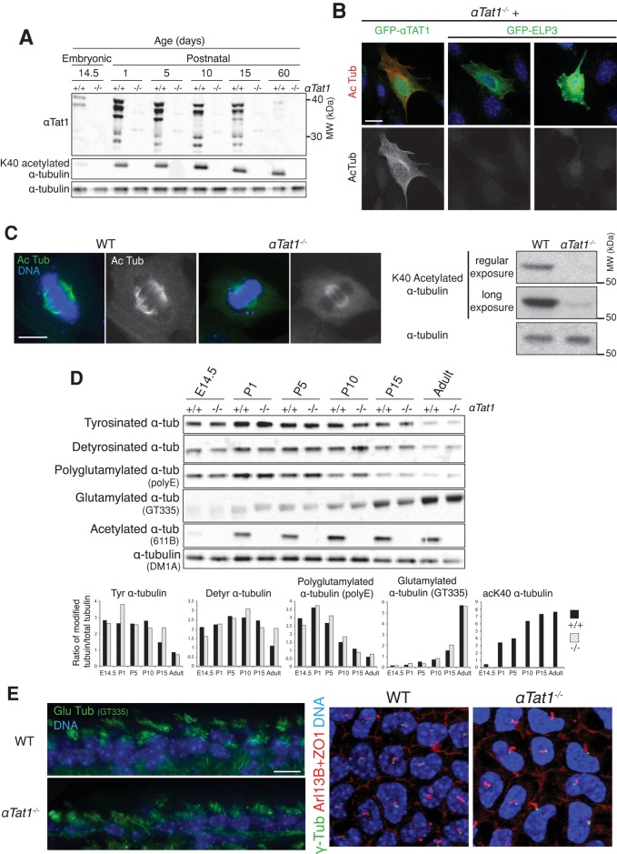
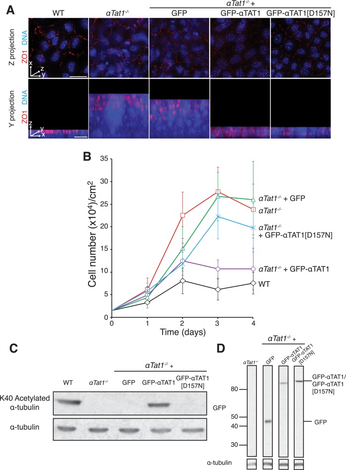
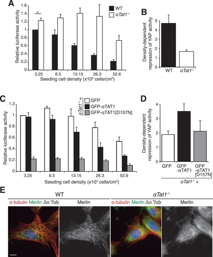
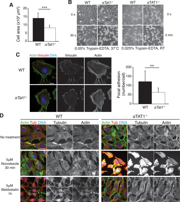
Acetylation of α-tubulin on lysine 40 marks long-lived microtubules in structures such as axons and cilia, and yet the physiological role of α-tubulin K40 acetylation is elusive. Although genetic ablation of the α-tubulin K40 acetyltransferase αTat1 in mice did not lead to detectable phenotypes in the developing animals, contact inhibition of proliferation and cell-substrate adhesion were significantly compromised in cultured αTat1(-/-) fibroblasts. First, αTat1(-/-) fibroblasts kept proliferating beyond the confluent monolayer stage. Congruently, αTat1(-/-) cells failed to activate Hippo signaling in response to increased cell density, and the microtubule association of the Hippo regulator Merlin was disrupted. Second, αTat1(-/-) cells contained very few focal adhesions, and their ability to adhere to growth surfaces was greatly impaired. Whereas the catalytic activity of αTAT1 was dispensable for monolayer formation, it was necessary for cell adhesion and restrained cell proliferation and activation of the Hippo pathway at elevated cell density. Because α-tubulin K40 acetylation is largely eliminated by deletion of αTAT1, we propose that acetylated microtubules regulate contact inhibition of proliferation through the Hippo pathway.


Figures

FIGURE 1:
FIGURE 1:
αTat1 is the major α-tubulin K40 acetyltransferase in vivo and is dispensable for mammalian CNS development and ciliogenesis. (A) Brain lysates from various developmental stages (E14.5, embryonic day 14.5; P1–P15, postnatal days 1–15) were immunoblotted for the indicated proteins. (B) αTat1−/− MEFs were transfected with GFP-αTat1 or GFP-ELP3 (green) and immunostained for GFP (green) and K40 acetylated α-tubulin (red). (C) Left, K40 acetylated α-tubulin immunostaining (green) of αTat1+/+ and αTat1−/− MEFs. Right, αTat1+/+ and αTat1−/− MEFs lysates were immunoblotted for K40 acetylated and total α-tubulin. (D) Brain lysates from αTat1+/+ and αTat1−/− mice at various developmental stages were immunoblotted for various α-tubulin posttranslational modifications. The quantitation of the immunoblots showed no major differences between wild-type and knockout mice. (E) Left, adult brain cryosections were stained with polyglutamylated α-tubulin (GT335 antibody, ependymal motile cilia, green). Right, basal bodies (γ-tubulin, green), primary cilia (Arl13B, red) and the cell–cell junction (ZO-1, red) were labeled in P6 corneal endothelium whole mounts. No defects in motile or primary cilia presence were noted in αTat1−/− mice. Scale bars: B, 20 μm; C, 10 μm; E, 10 μm.
FIGURE 2:
FIGURE 2:
αTat1 is required for contact inhibition of proliferation. (A) αTat1+/+, αTat1−/−, and αTat1−/− cells expressing GFP, GFP-αTAT1, or GFP-αTAT1[D157N] were seeded at 15,000 cells/cm2. Five days after seeding, cells were fixed and stained for the cell–cell junction marker ZO-1 (red) and DNA (blue). Wild-type cells form a monolayer, seen as clearly separated nuclei in z-projection and as a row of evenly distributed nuclei in the projection along the y-axis. Expression of GFP-αTAT1 or GFP-αTAT1[D157N] restored monolayer formation, whereas GFP alone did not. In addition, the pattern of ZO1 staining suggests that the bottom layers of the αTat1−/− cultures have lost polarity, whereas the top layer does retain some level of polarity. In addition, the rescue of polarity by expression of GFP-αTAT1 appears to only be partial. (B) αTat1+/+, αTat1−/−, and αTat1−/− cells expressing GFP, GFP-αTAT1, or GFP-αTAT1[D157N] were seeded at 15,000 cells/cm2, counted every day, and fed every other day. After 2 d of culture, wild-type cells reached a stereotypical saturating density (∼60,000 cells/cm2) and stayed quiescent for the rest of the assay. αTat1−/− cells bypassed the normal saturating density soon after day 1 and reached a saturating density fourfold higher than wild type after 3 d of culture. GFP-αTAT1 expression in αTat1−/− cells restored the saturating density to near-wild-type levels, whereas GFP or GFP-αTAT1[D157N] did not. (C) Lysates of αTat1+/+, αTat1−/−, and αTat1−/− cells expressing GFP, GFP-αTAT1, or GFP-αTAT1[D157N] were immunoblotted for K40 acetylated α-tubulin. Only GFP-αTAT1 expression restores K40 α-tubulin acetylation to wild-type levels. (D) Cell lysates from αTat1−/− and αTat1−/− cells expressing GFP, GFP-αTAT1, or GFP-αTAT1[D157N] were immunoblotted for GFP and α-tubulin. αTat1−/− cell lines expressing GFP and GFP-αTAT1[D157N] were selected to match GFP-αTAT1 expression levels. All lanes come from the same membrane and, as such, have the same exposure. Scale bars: A, 30 μm (top), 5 μm (bottom).
FIGURE 3:
FIGURE 3:
αTat1 is essential for proper Hippo signaling (A) YAP/TAZ luciferase assay (8XGTIIC-luc) in wild-type (WT) and αTat1−/− cells seeded at increasing densities. Whereas YAP/TAZ activity is down-regulated with increasing cell density in WT cells, knockout cells exhibit a high luciferase signal even at the highest cell density. Relative luciferase activity was normalized to the value of WT cells at the sparsest density. (B) Ratio of the relative luciferase activity for the lowest over the highest cell density in WT and αTat1−/− cells. This ratio reflects the cell's ability to down-regulate YAP/TAZ activity upon increasing cell density. (C) YAP/TAZ luciferase assay (8XGTIIC-luc) in αTat1−/− cells stably expressing GFP, GFP-αTat1, and GFP-αTAT1[D157N], seeded at increasing densities. Only GFP-αTAT1[D157N] rescues the unusually high levels of YAP/TAZ activity in αTat1−/− cells. (D) Ratio of the relative luciferase activity for the lowest over the highest cell density in αTat1−/− cells stably expressing GFP, GFP-αTat1, and GFP-αTAT1[D157N]. Only GFP-αTat1 was able to rescue YAP/TAZ activity down-regulation upon cell density increase in αTat1−/− cells. (E) WT and αTat1−/− cells were immunolabeled for Merlin and K40 acetylated and total α-tubulin. Whereas in WT cells merlin was localized to particles along the microtubules, it exhibited diffuse localization in αTat1−/− cells. Scale bar, 10 μm.
FIGURE 4:
FIGURE 4:
αTat1 is required for cell adhesion. (A) The surface area of αTat1+/+ and αTat1−/− cells was measured using ImageJ (n = 10 cells/genotype). Knockout cells have a very significant decrease in cell area compared with wild-type cells (p = 7.5 × 10⁻⁵). (B) αTat1+/+ and αTat1−/− cells were trypsinized with 37ºC, 0.05% trypsin-EDTA or 0.025%, room temperature trypsin-EDTA. At the time points shown, trypsin-EDTA has little effect on wild-type cells, yet it rounds up most αTat1−/− cells. (C) αTat1+/+ and αTat1−/− cells were labeled with an anti-vinculin antibody (red) and Alexa 488–phalloidin (green). The αTat1−/− cells showed reduced number of stress fibers and disorganized actin cytoskeleton. Using ImageJ, we quantified the number of focal adhesions per cell and found that it decreased in αTat1−/− cells compared with wild-type (p = 0.01, n = 10 cells/genotype). (D) αTat1+/+ and αTat1−/− cells were treated with nocodazole or blebbistatin and immunostained for α-tubulin (red). Actin was labeled using Alexa 488–phalloidin (green). Whereas nocodazole treatment stimulates stress fiber formation in wild-type cells, no or few stress fibers were observed in αTat1−/− cells, which rounded up and detached. In blebbistatin-treated wild-type cells, the inhibition of myosin II function induces cytoplasmic collapse. In αTat1−/− cells, blebbistatin treatment did not yield any major morphological alteration. See Supplemental Figure S5 for a shorter exposure, where the fluorescence levels of αTat1−/− cells treated with nocodazole are not saturated. The apparent increase in tubulin and actin staining in αTat1−/− cells treated with nocodazole is likely due the rounding up of cells, which will generate a significant amount of out-of-focus fluorescence captured by epifluorescence. **p ≤ 0.01, ***p ≤ 0.001, two-sided Wilcoxon test. Scale bars: B, 50 μm (left), 100 μm (right); C, 10 μm; D, 15 μm.

References

    1. Abercrombie M. Contact-dependent behavior of normal cells and the possible significance of surface changes in virus-induced transformation. Cold Spring Harb Symp Quant Biol. 1962;27:427–431. - PubMed
    1. Abercrombie M. Contact inhibition and malignancy. Nature. 1979;281:259–262. - PubMed
    1. Akella JS, Wloga D, Kim J, Starostina NG, Lyons-Abbott S, Morrissette NS, Dougan ST, Kipreos ET, Gaertig J. MEC-17 is an alpha-tubulin acetyltransferase. Nature. 2010;467:218–222. - PMC - PubMed
    1. Aragona M, Panciera T, Manfrin A, Giulitti S, Michielin F, Elvassore N, Dupont S, Piccolo S. A mechanical checkpoint controls multicellular growth through YAP/TAZ regulation by actin-processing factors. Cell. 2013;154:1047–1059. - PubMed
    1. Artemenko Y, Batsios P, Borleis J, Gagnon Z, Lee J, Rohlfs M, Sanseau D, Willard SS, Schleicher M, Devreotes PN. Tumor suppressor Hippo/MST1 kinase mediates chemotaxis by regulating spreading and adhesion. Proc Natl Acad Sci USA. 2012;109:13632–13637. - PMC - PubMed

Publication types

MeSH terms

LinkOut - more resources