. 2014 Apr 17;10(4):e1004074.
doi: 10.1371/journal.ppat.1004074. eCollection 2014 Apr.

The apical complex provides a regulated gateway for secretion of invasion factors in Toxoplasma

Affiliations

The apical complex provides a regulated gateway for secretion of invasion factors in Toxoplasma

Nicholas J Katris et al. PLoS Pathog. .

Abstract

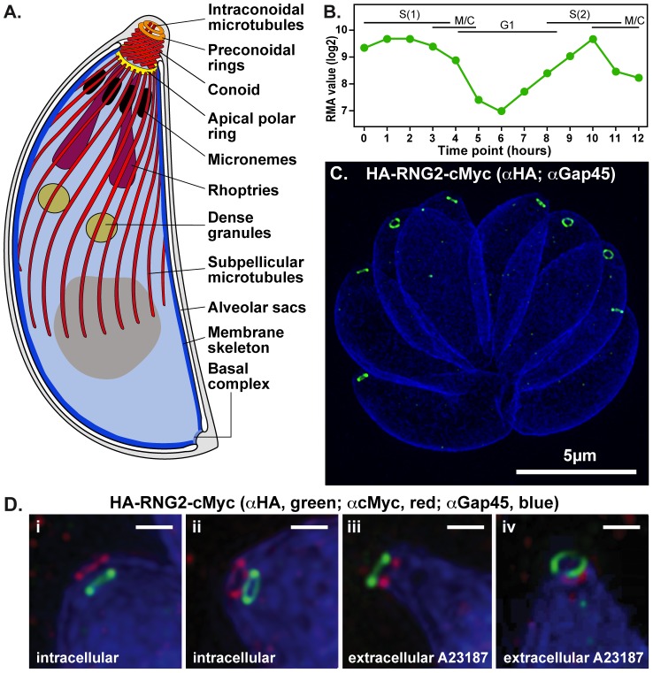
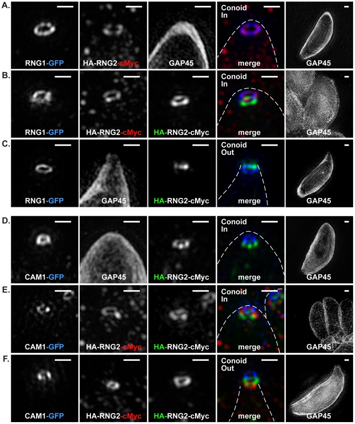
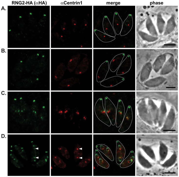
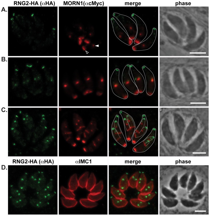
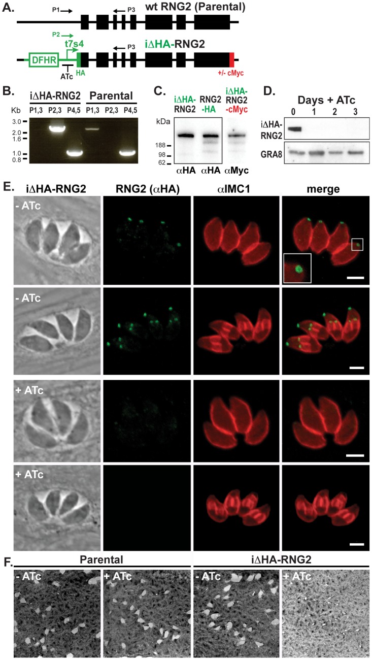
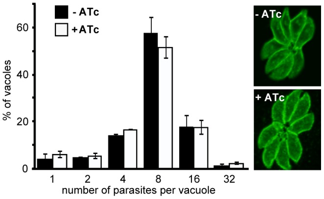
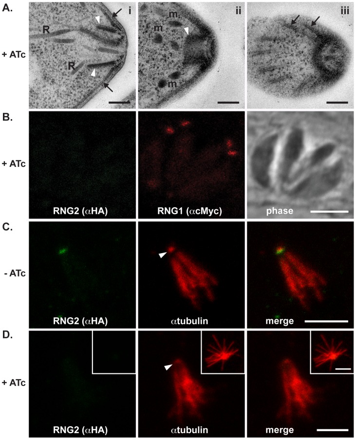
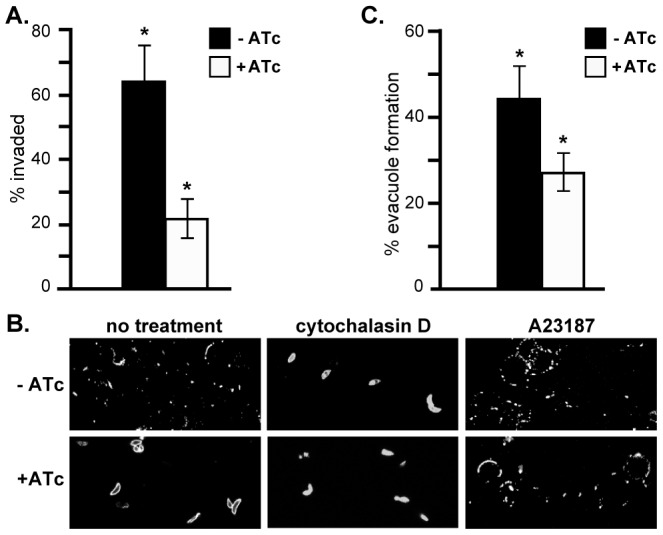
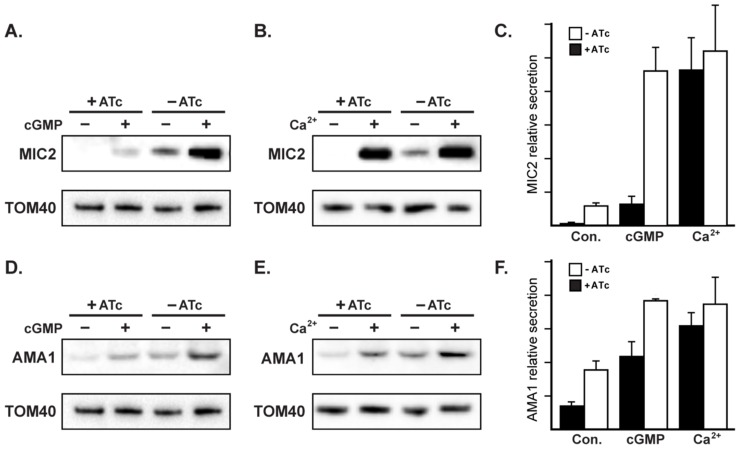
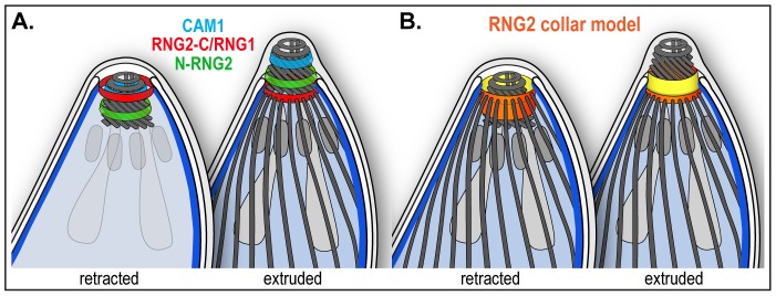
The apical complex is the definitive cell structure of phylum Apicomplexa, and is the focus of the events of host cell penetration and the establishment of intracellular parasitism. Despite the importance of this structure, its molecular composition is relatively poorly known and few studies have experimentally tested its functions. We have characterized a novel Toxoplasma gondii protein, RNG2, that is located at the apical polar ring--the common structural element of apical complexes. During cell division, RNG2 is first recruited to centrosomes immediately after their duplication, confirming that assembly of the new apical complex commences as one of the earliest events of cell replication. RNG2 subsequently forms a ring, with the carboxy- and amino-termini anchored to the apical polar ring and mobile conoid, respectively, linking these two structures. Super-resolution microscopy resolves these two termini, and reveals that RNG2 orientation flips during invasion when the conoid is extruded. Inducible knockdown of RNG2 strongly inhibits host cell invasion. Consistent with this, secretion of micronemes is prevented in the absence of RNG2. This block, however, can be fully or partially overcome by exogenous stimulation of calcium or cGMP signaling pathways, respectively, implicating the apical complex directly in these signaling events. RNG2 demonstrates for the first time a role for the apical complex in controlling secretion of invasion factors in this important group of parasites.


Conflict of interest statement

The authors have declared that no competing interests exist.

Figures

Figure 1
Figure 1. RNG2 apical rings.
(A) Schematic of Toxoplasma gondii cell representing the structural elements of the apical complex, the cell pellicle, and the secretory organelles. The conoid is shown extruded. (B) RNG2 expression throughout the cell cycle based on dataset of Behnke et al. . (C) 3D-SIM image of intracellular HA-RNG2-cMyc parasites showing the RNG2 N-terminus (HA, green) against GAP45 labeling of the IMC (blue). (D). 3D-SIM images of intracellular parasites with retracted conoids (i, ii) and extracellular parasites treated with A23187 to extrude conoids (iii, iv) labeled for RNG2 N-terminus (green) and C-terminus (red), and GAP45 (blue). Scale bar = 500 nm.
Figure 2
Figure 2. 3D-SIM of RNG2 location relative apical polar ring marker RNG1 and conoid marker CAM1.
(A-C) RNG1-GFP (pseudo-colored blue), and (D-F) CAM1-GFP (pseudo-colored blue) colocalized with HA (green) and cMyc (red) of the HA-RNG2-cMyc fusion. Conoid position is indicated. Individual immuno-signals are shown in monochrome, and colored in the merged image where GAP45 labeling is shown by a dashed outline. Right hand column shows GAP45 labeling of each cell at 0.25 magnification. Scale bars = 500 nm.
Figure 3
Figure 3. RNG2 appears in daughter cells after centrosome duplication.
RNG2-HA (green) cells immuno-labeled for centrin1 (red) show single centrosome duplication at the beginning of daughter cell formation (A, B), after which RNG2 appears in association with each centrosome (C). (D) As centrosome pairs separate, RNG2 dissociates and forms rings, but leaves a trace of RNG2 in association with the centrosome (e.g. arrowheads). Inferred daughter buds shown with dashed lines, scale bars = 3 μm.
Figure 4
Figure 4. RNG2 appears before centrocone duplication or IMC1 association with daughter pellicles.
(A, B, C) RNG2-HA (green) co-expressed with MORN1-cMyc (red). MORN1 can be seen at the basal complex of both mother (e.g. A, open arrowhead) and daughter cell pellicles, and at the centrocone (e.g. A, filled arrowhead). Inferred daughter buds shown with dashed lines (C) Elongation of the centrocone occurs prior to resolution of two centrocones and mitosis. (D) Immuno-labeled RNG2-HA (green) and IMC1 (red). Scale bars = 3 μm.
Figure 5
Figure 5. RNG2 is required for parasite growth.
(A) Schematic of the chromosomal locus of wild type RNG2 and the insertional mutant iΔHA-RNG2 showing the tetracycline regulatable promoter (t7s4), N-terminal HA tag, C-terminal cMyc tag (integrated separately) selectable marker DHFR, exon structure of RNG2 (black boxes), and primers (P1–3) used to verify correct integration. (B) PCR analysis of genomic rng2 locus after integration of the iΔHA insert. Primer P1–3 locations shown in (A) while P4,5 amplify tic22 as a positive control. (C) Western blot of three versions of tagged RNG2 using either HA or cMyc antibodies (equal protein loading in all lanes). (D) Western blot of HA-RNG2 expression after 0 to 3 days of anhydrotetracycline (ATc) treatment. Dense granule protein GRA8 used as a loading control. (E) Immuno-fluorescence detection of HA-RNG2 (green) and cell pellicles (IMC1, red) after no or one day of ATc treatment. Scale bar = 3 μm. (F) Plaque assay measuring parasite growth over 8 days of either the parental or knockdown cell lines, in the absence (−) or presence (+) of ATc.
Figure 6
Figure 6. RNG2 is not required for intracellular parasite replication.
Replication rate of iΔHA-RNG2 parasites grown with (+) or without (−) ATc treatment measured by parasite number per parasitophorous vacuole after 24 hours of growth post infection. >200 vacuoles were counted for each of three biological replicates, error bars = standard error of the mean. Representative SAG1-stained vacuoles show no gross difference between + or − ATc-treated cells.
Figure 7
Figure 7. RNG2 knockdown results in no obvious structural change.
(A) Transmission electron microscopy of ATc-treated parasites show typical apical structures. (i) Transverse section of apical complex showing rhoptries (R) extending within the conoid (white arrowheads) and microtubules and the IMC (arrows) converging on the apical polar ring. (ii) Oblique section of the apical complex shows the retracted conoid (arrowhead), preconoidal rings and micronemes (m), while a glancing section shows subpellicular microtubules (arrows) converging on the apical polar ring (iii).(B) iΔHA-RNG2 cells treated with ATc and co-expressing RNG1-cMyc show no change in RNG1 association with the apical polar ring. (C) Detergent-extracted pellicles show HA-RNG2 persists with the microtubular conoid (arrowhead) and sub-pellicular basket. (D) With ATc treatment and RNG2 knockdown these extracted pellicles remain unchanged with microtubules attached to the conoid (arrowhead). Inset image shows a splayed microtubular basket. Black scale bar = 200 nm, white scale bar = 3 μm.
Figure 8
Figure 8. RNG2 is required for invasion, motility and rhoptry evacuole formation.
(A) Percentage parasite invasion success of iΔHA-RNG2 cells grown either with or without ATc. >200 parasites were scored for each of three biological replicates. (B) Gliding motility detected by SAG1-positive trails of iΔHA-RNG2 cells with or without ATc. Motility was also assessed with further treatments of actin polymerization inhibitor cytochalasin D, or calcium ionophore A23187. (C) Percentage of ROP1-positive evacuoles formed by iΔHA-RNG2 cells grown with or without ATc. >200 parasites were scored for each of six biological replicates. Error bars  =  standard error of the mean, * denotes significant differences (P<0.05, two-tailed Student's t-test).
Figure 9
Figure 9. RNG2 has a role in regulated microneme secretion.
Constitutive secretion of microneme proteins MIC2 (A, B) and AMA1 (D, E) from extracellular iΔHA-RNG2 cells grown with or without ATc. Secretion was also assessed with cGMP stimulation by Zaprinast (A, D), or with calcium stimulation by A23187 (B, E). Tom40 detection in cell pellets provides a control for parasite number used for secretion assays. (C, F) Secretion averages from biological replicates (n = 6 for constitutive (Con.) secretion; n = 4 for stimulated secretion). Error bars  =  standard error of the mean.
Figure 10
Figure 10. Schematic of RNG2 location within the apical complex.
(A) Inferred positions of the N and C termini of RNG2 (labeled N-RNG2 and RNG2-C, respectively), conoid marker CAM1, and apical polar ring marker RNG1 within the structures of the apical complex, and with the conoid either retracted (subpellicular microtubules removed) or extruded. (B) Spatial model of RNG2 (orange and yellow), based on locations of protein termini, forming a collar between the apical polar ring and conoid. The collar is inverted upon conoid extrusion, potentially turning inside-out. See Figure 1 for full labeling of structures.

References

    1. Adl SM, Leander BS, Simpson AGB, Archibald JM, Andersen OR, et al. (2007) Diversity, Nomenclature, and Taxonomy of Protists. Systematic Biol 56: 684–689. - PubMed
    1. WHO (2012) World Malaria Report 2012.: Geneva, Switzerland: World Health Organisation.
    1. Montoya JG, Liesenfeld O (2004) Toxoplasmosis. Lancet 363: 1965–1976. - PubMed
    1. Leander BS (2008) Marine gregarines: evolutionary prelude to the apicomplexan radiation? Trends Parasitol 24: 60–67. - PubMed
    1. Simdyanov TG, Kuvardina ON (2007) Fine structure and putative feeding mechanism of the archigregarine Selenidium orientale (Apicomplexa: Gregarinomorpha). Eur J Protistol 43: 17–25. - PubMed

Publication types