Randomized Controlled Trial
. 2014 Apr 17;10(4):e1004079.
doi: 10.1371/journal.ppat.1004079. eCollection 2014 Apr.

Exposure-dependent control of malaria-induced inflammation in children

Affiliations
Randomized Controlled Trial

Exposure-dependent control of malaria-induced inflammation in children

Silvia Portugal et al. PLoS Pathog. .

Abstract

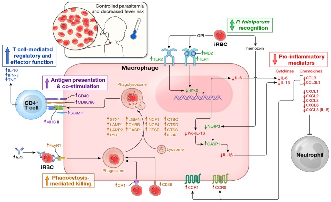
In malaria-naïve individuals, Plasmodium falciparum infection results in high levels of parasite-infected red blood cells (iRBCs) that trigger systemic inflammation and fever. Conversely, individuals in endemic areas who are repeatedly infected are often asymptomatic and have low levels of iRBCs, even young children. We hypothesized that febrile malaria alters the immune system such that P. falciparum re-exposure results in reduced production of pro-inflammatory cytokines/chemokines and enhanced anti-parasite effector responses compared to responses induced before malaria. To test this hypothesis we used a systems biology approach to analyze PBMCs sampled from healthy children before the six-month malaria season and the same children seven days after treatment of their first febrile malaria episode of the ensuing season. PBMCs were stimulated with iRBC in vitro and various immune parameters were measured. Before the malaria season, children's immune cells responded to iRBCs by producing pro-inflammatory mediators such as IL-1β, IL-6 and IL-8. Following malaria there was a marked shift in the response to iRBCs with the same children's immune cells producing lower levels of pro-inflammatory cytokines and higher levels of anti-inflammatory cytokines (IL-10, TGF-β). In addition, molecules involved in phagocytosis and activation of adaptive immunity were upregulated after malaria as compared to before. This shift was accompanied by an increase in P. falciparum-specific CD4+Foxp3- T cells that co-produce IL-10, IFN-γ and TNF; however, after the subsequent six-month dry season, a period of markedly reduced malaria transmission, P. falciparum-inducible IL-10 production remained partially upregulated only in children with persistent asymptomatic infections. These findings suggest that in the face of P. falciparum re-exposure, children acquire exposure-dependent P. falciparum-specific immunoregulatory responses that dampen pathogenic inflammation while enhancing anti-parasite effector mechanisms. These data provide mechanistic insight into the observation that P. falciparum-infected children in endemic areas are often afebrile and tend to control parasite replication.


Conflict of interest statement

The authors have declared that no competing interests exist.

Figures

Figure 1
Figure 1. A molecular pattern of restrained inflammation and enhanced anti-parasite effector function upon P. falciparum re-exposure.
(A) PBMCs were collected from 34 healthy children with blood smears negative for P. falciparum infection before the malaria season (HB) and 7 days after treatment of their first febrile malaria episode of the ensuing malaria season when malaria symptoms had resolved (d7). RNA was extracted from PBMCs immediately after thawing and hybridized onto Affymetrix GeneChip Human 1.0 ST arrays. RNA from all 68 PBMC samples was of sufficient quantity and quality for microarray analysis. Nine of 68 samples did not pass the microarray quality assessment and were removed from further analysis (see Supplemental Figure 1A) such that 25 children with paired RNA samples at the healthy baseline and 7 days after malaria were analyzed. The heat map shows ex vivo RMA-normalized log2 ratios (d7/HB) of differentially expressed genes (rows) for each child (columns). Genes are grouped and color-coded by function as indicated. (B) PBMCs analyzed by FACS for B cells (CD19+), T cells (CD3+), CD3+CD4+ T cells, CD3+CD8+ T cells, and monocytes (CD14+) at the healthy baseline and after malaria. (n = 34 children; except CD14+ monocytes, n = 30). (C) Ratio of monocyte percentage (d7/HB) versus the ratio of the expression level of monocyte-derived pro-inflammatory cytokines and chemokines (d7/HB). Each point represents an individual subject (n = 21 children with paired samples). (D) RNA was extracted from PBMCs of the same 34 children after 18 h of in vitro stimulation with P. falciparum-infected red blood cell (iRBC) lysate. After stimulation with iRBC lysate, 22 of the 34 children had RNA samples from both time points of sufficient quantity and quality for microarray analysis and also passed the microarray quality assessment. The heat map shows RMA-normalized log2 ratios (d7/HB) of differentially expressed genes (rows) for each child (columns) in response to in vitro iRBC lysate stimulation. Genes are grouped and color-coded by function as indicated. (E) q-RT-PCR confirmation of the microarray data. The data represent the results of one experiment with 6 genes (IL1B, IL6, IL10, TGFB1, TLR2, CXCL5) from 17 subjects at two time points (d7 and HB) from both the ex vivo unstimulated and in vitro iRBC-stimulated datasets. Each symbol represents a single gene at a given time point. PCR expression computed as antilog2 –dCT. n = 497 XY pairs. (F) q-RT-PCR expression of genes encoding the pro-inflammatory cytokines IL1-β and IL-6 and the anti-inflammatory cytokine TGF-β in PBMCs of children (n = 17) collected at the healthy baseline (HB) and after resolution of febrile malaria (d7), either directly ex vivo (unstimulated) or after in vitro stimulation with iRBCs for 18 h. ns, not significant (P≥0.05), P values determined by the paired ttest (B), Pearson's (C), Spearman's (E) or paired Wilcoxon rank sum test (F). Data are shown as the means ± s.d. (B) or means ± s.e.m. (F).
Figure 2
Figure 2. P. falciparum-inducible IL-10 production is upregulated upon re-exposure and partially maintained by persistent asymptomatic infection.
(A) Production of IL-10, IL-8, IL1-β, IL-6 and TNF by PBMCs in response to in vitro stimulation with iRBC lysate at the healthy baseline before the malaria season (HB) and 7 days after malaria (d7) (n = 28 children with paired samples). (B) Production of IL-1β and IL-6 by isolated monocytes/macrophages after 6 h of in vitro stimulation with iRBC lysate. Results are shown as the ratio of cytokines produced 14 days after malaria (d14) versus the healthy baseline before the malaria season (HB) (n = 9 children with paired samples) (P = 0.0066 and P = 0.0003 for IL-1β and IL-6 respectively). (C) A positive control showing IL-10 production by PBMCs in response to in vitro stimulation with iRBC lysate in the presence of blocking antibodies specific for IL-10 and the IL-10 receptor or the isotype control (n = 17). (D,E) TNF and IL-6 production by PBMCs in response to in vitro stimulation with iRBC lysate in the presence of blocking antibodies specific for IL-10 and the IL-10 receptor at the healthy baseline (HB), 7 days after malaria (d7) and at the healthy baseline after the subsequent 6-month dry season (HB′) (n = 20 children, 9 paired in the 3 conditions). (F) IL-10 production by PBMCs in response to in vitro stimulation with iRBC lysate at the healthy baseline before the malaria season (HB), 7 days after malaria (d7) and at the healthy baseline after the subsequent 6-month dry season (HB′), a period of little to no P. falciparum transmission (n = 15 children). (G) IL-10 production by PBMCs in response to in vitro stimulation with iRBC lysate among children with asymptomatic P. falciparum infection at the end of the dry season (HB Pf+, n = 16) versus aged-matched, healthy uninfected children at the same time point (HB Pf−, n = 34). Data are presented as fold change relative to PBMCs stimulated with uninfected RBC (uRBC) lysate (A, F, G). ns, not significant (P≥0.05), P values were determined by a paired ttest (A, C), one-sample Student's T-tests comparing the mean ratio against a 1∶1 ratio (B), paired ttest followed by Bonferroni's test (D–F) or unpaired ttest (G). Data are shown as the means ± s.d.
Figure 3
Figure 3. P. falciparum-inducible IL-10 is mainly produced by CD4+CD25+Foxp3 T cells that co-produce IFNγ and TNF.
(A) PBMCs from the healthy baseline (HB), 7 days after malaria (d7), and at the healthy baseline at the end of the subsequent dry-season (HB′) were stimulated for 18 h with iRBC lysate and assayed for the production of IL-10, IFNγ and TNF by intra-cellular FACS. Results are shown as the ratio of live CD3+ CD4+ antigen-experienced cells (CD45RO+ CD27+, CD45RO+ CD27, and CD45RO CD27) producing IL-10, IFN-γ or TNF in response to stimulation with iRBC lysate vs. uninfected RBC (uRBC) lysate (n = 16, 13 paired samples). (B) Overlay of IL-10-producing cells (red) among all live cells (gray) in a CD3 vs. CD4 dot plot (top) (n = 14), and IL-10-producing CD4+ T cells (red) with all CD4+ T cells (gray) in CD25 vs. FoxP3 dot plot (bottom) (n = 9; representative subject shown). (C) Using SPICE analysis, cytokine-producing CD4+ T cells were divided into 7 distinct subpopulations producing any combination of IL-10, IFNγ and TNF (n = 16). (D) Pie chart representation of the combination of cytokines produced by CD4+ T cells after iRBC stimulation for 3 representative donors 7 days after malaria (d7). The black arcs indicate the IL-10-producing CD4+ T cells. (E) Representative FACS plots of live CD3+ CD4+ antigen-experienced cells producing IL-10, IFNγ and TNF after iRBC stimulation of PBMCs collected at the healthy baseline (HB), 7 days after malaria (d7) and at the healthy baseline at the end of the subsequent dry-season (HB′). (F) CD4+ T cells were isolated from PBMCs which had been collected from children 7 days after malaria and were then stimulated for 18 h with iRBC or uRBC lysate in the absence (CD4+T d7) or presence of non-CD4+T cells isolated from PBMCs of the same individuals collected at either the healthy baseline (CD4+T d7 + nonCD4+T HB) or 7 days after malaria (CD4+T d7 + nonCD4+T d7) (n = 8 paired samples). (G) PBMCs collected from children 7 days after malaria were stimulated for 18 h with iRBC lysate and assayed for the production of IL-10 in the presence (αMHC-II) or absence (isotype) of antibodies specific for HLA-DR, -DQ and -DP (n = 8). ns, not significant (P≥0.05), P values determined by a linear mixed model for repeated measures ANOVA with Tukey HSD post hoc tests (A) and permutation re-sampling tests (F, G). Data are shown as the means ± s.d.
Figure 4
Figure 4. Proposed model by which children remain asymptomatic and control parasitemia upon P. falciparum re-exposure.
In children without prior or recent malaria exposure, P. falciparum infection induces a robust pro-inflammatory cytokine and chemokine response (e.g. IL-1β, IL-6, IL-8) whereas effector mechanisms that mediate parasite clearance (phagocytosis, phagolysosome activation, antigen presentation, T cell co-stimulation and IFN-γ production by CD4+ T cells) are not readily inducible, leaving children susceptible to fever and other systemic symptoms of malaria as well as poorly controlled parasite replication. In contrast, febrile malaria induces an exposure-dependent regulatory state (shown here) whereby re-exposure to P. falciparum results in reduced production of pro-inflammatory cytokines and chemokines and enhanced expression of regulatory cytokines (e.g. IL-10 production by CD4+ T cells) and pathways involved in phagocytosis-mediated clearance of infected red blood cells and activation of adaptive immunity, thus enabling children to remain asymptomatic and control parasite replication in the face of ongoing P. falciparum exposure. In addition, P. falciparum-specific IgG levels are low in children who have not been recently exposed to malaria, but transiently increase in response to P. falciparum infection , , further enhancing exposure-dependent parasite clearance through opsonization and phagocytosis of infected erythrocytes. Arrows indicate the direction of expression observed in this study of molecules at the mRNA and/or protein levels induced by P. falciparum re-exposure after febrile malaria relative to responses induced by P. falciparum exposure at the healthy baseline. Molecules are color-coded by biological function.

References

    1. Collins WE, Jeffery GM (1999) A retrospective examination of sporozoite- and trophozoite-induced infections with Plasmodium falciparum: development of parasitologic and clinical immunity during primary infection. Am J Trop Med Hyg 61: 4–19. - PubMed
    1. Day NP, Hien TT, Schollaardt T, Loc PP, Chuong LV, et al. (1999) The prognostic and pathophysiologic role of pro- and antiinflammatory cytokines in severe malaria. J Infect Dis 180: 1288–1297. - PubMed
    1. Lyke KE, Burges R, Cissoko Y, Sangare L, Dao M, et al. (2004) Serum levels of the proinflammatory cytokines interleukin-1 beta (IL-1beta), IL-6, IL-8, IL-10, tumor necrosis factor alpha, and IL-12(p70) in Malian children with severe Plasmodium falciparum malaria and matched uncomplicated malaria or healthy controls. Infect Immun 72: 5630–5637. - PMC - PubMed
    1. Walther M, Woodruff J, Edele F, Jeffries D, Tongren JE, et al. (2006) Innate immune responses to human malaria: heterogeneous cytokine responses to blood-stage Plasmodium falciparum correlate with parasitological and clinical outcomes. J Immunol 177: 5736–5745. - PubMed
    1. Urban BC, Ing R, Stevenson MM (2005) Early interactions between blood-stage plasmodium parasites and the immune system. Curr Top Microbiol Immunol 297: 25–70. - PubMed

Publication types