. 2014 Jun;42(10):6726-41.
doi: 10.1093/nar/gku269. Epub 2014 Apr 17.

DNA-protein π-interactions in nature: abundance, structure, composition and strength of contacts between aromatic amino acids and DNA nucleobases or deoxyribose sugar

Affiliations

DNA-protein π-interactions in nature: abundance, structure, composition and strength of contacts between aromatic amino acids and DNA nucleobases or deoxyribose sugar

Katie A Wilson et al. Nucleic Acids Res. 2014 Jun.

Abstract

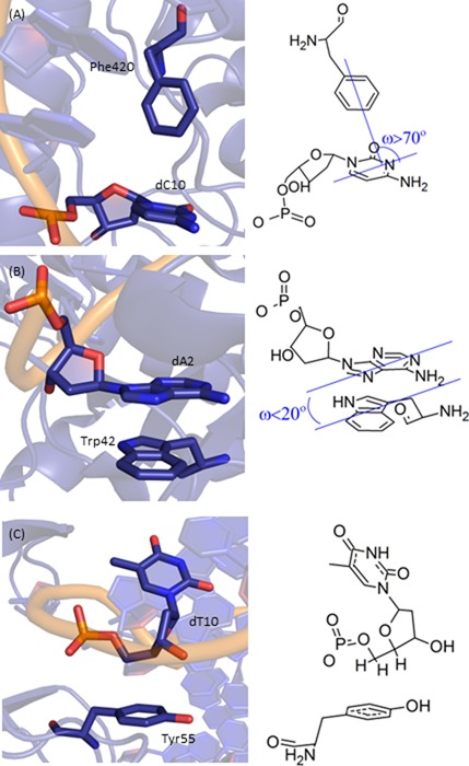
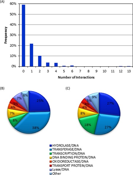
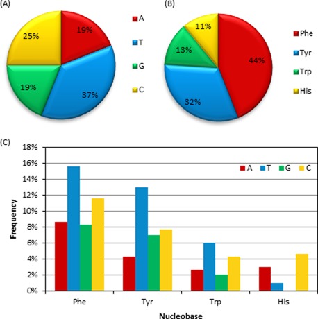
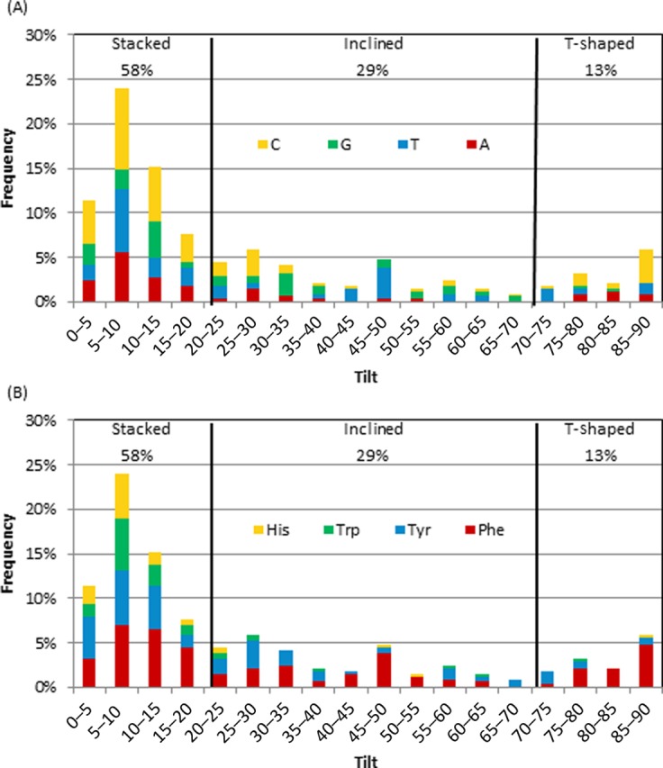
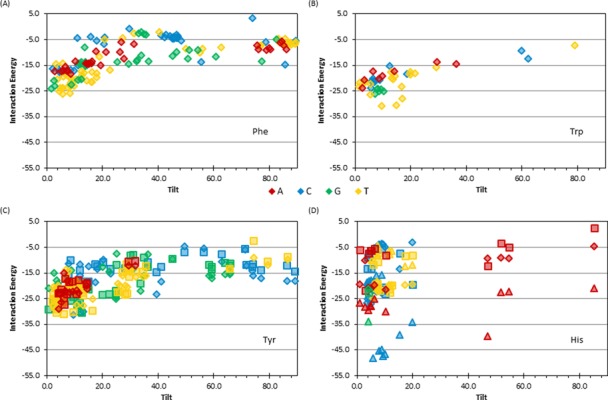
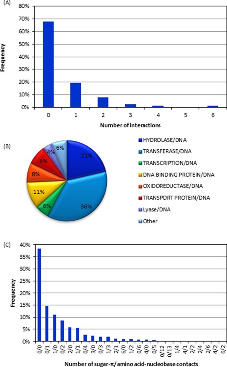
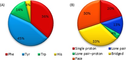
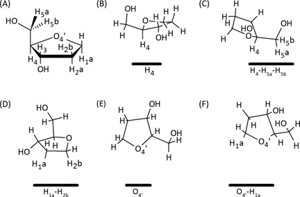
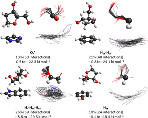
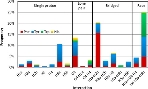
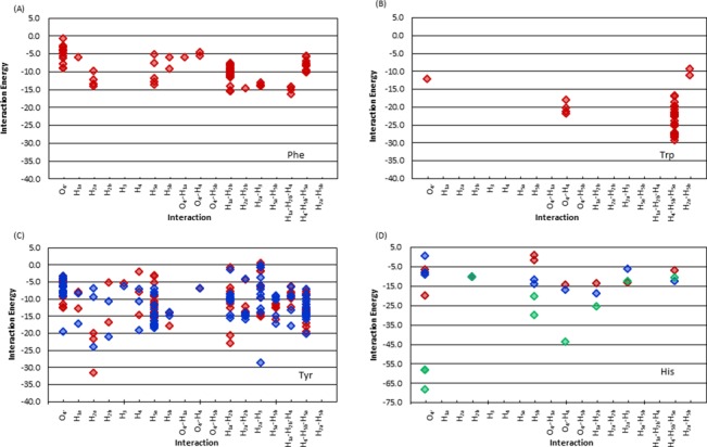
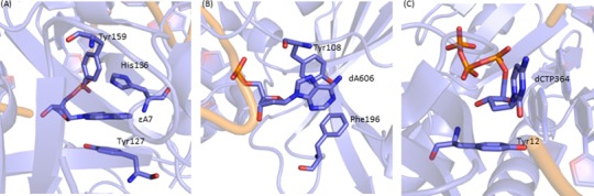
Four hundred twenty-eight high-resolution DNA-protein complexes were chosen for a bioinformatics study. Although 164 crystal structures (38% of those searched) contained no interactions, 574 discrete π-contacts between the aromatic amino acids and the DNA nucleobases or deoxyribose were identified using strict criteria, including visual inspection. The abundance and structure of the interactions were determined by unequivocally classifying the contacts as either π-π stacking, π-π T-shaped or sugar-π contacts. Three hundred forty-four nucleobase-amino acid π-π contacts (60% of all interactions identified) were identified in 175 of the crystal structures searched. Unprecedented in the literature, 230 DNA-protein sugar-π contacts (40% of all interactions identified) were identified in 137 crystal structures, which involve C-H···π and/or lone-pair···π interactions, contain any amino acid and can be classified according to sugar atoms involved. Both π-π and sugar-π interactions display a range of relative monomer orientations and therefore interaction energies (up to -50 (-70) kJ mol(-1) for neutral (charged) interactions as determined using quantum chemical calculations). In general, DNA-protein π-interactions are more prevalent than perhaps currently accepted and the role of such interactions in many biological processes may yet to be uncovered.


Figures

Figure 1.
Figure 1.
Examples of (A) nucleobase–amino acid π–π T-shaped interaction (PDB ID: 2WQ7), (B) nucleobase–amino acid π–π stacking interaction (PDB ID: 3MR5) and (C) deoxyribose–amino acid sugar–π interaction (PDB ID: 3BKZ).
Figure 2.
Figure 2.
(A) Number of nucleobase–amino acid stacking/T-shaped interactions identified in PDB structures in the present study. (B) Types of proteins in which nucleobase–amino acid stacking/T-shaped interactions were found. (C) Overall composition of the proteins in the crystal structures considered in the present work.
Figure 3.
Figure 3.
The proportions of (A) nucleobases, (B) amino acids and (C) nucleobase–amino acid combinations in DNA–protein π–π stacked and T-shaped orientations found in nature.
Figure 4.
Figure 4.
Frequency of tilt angle (degrees) between the ring planes for all interactions according to the (A) nucleobase or (B) amino acid.
Figure 5.
Figure 5.
Binding energy of nucleobase–amino acid π–π interactions with respect to the tilt angle (degrees) for dimers involving (A) Phe, (B) Trp, (C) Tyr (for TyrCW (diamonds) and TyrCCW (squares) and (D) His (for Hisδ (diamonds), Hisϵ (squares) and His+ (triangles) (see Supplementary Figure S1, SI, for the definition of different Tyr and His conformations).
Figure 6.
Figure 6.
(A) The number of sugar–π contacts found in each structure. (B) Types of proteins in which sugar–π interactions were found. (C) The number of sugar–π and nucleobase–amino acid interactions observed in crystal structures considered in the present work.
Figure 7.
Figure 7.
(A) Composition of sugar–π interactions found in nature as a function of amino acid. (B) Frequency of sugar–π interactions found in nature with respect to the class of contact.
Figure 8.
Figure 8.
(A) Numbering scheme of the sugar moiety. Representative sugar–π interactions identified in crystal structures for (B) single proton, (C) face, (D) bridged, (E) lone pair and (F) lone pair–proton interactions (the amino acid is represented by a solid black line below the sugar).
Figure 9.
Figure 9.
Example dimer and overlay of all dimers for the four most common sugar–π contacts identified in crystal structures, including calculated binding strengths.
Figure 10.
Figure 10.
Frequency of sugar–π interactions found in nature with respect to the type of contact and the amino acid.
Figure 11.
Figure 11.
Binding energies of sugar–π interactions with respect to the type of contact for dimers involving (A) Phe, (B) Trp, (C) Tyr (for TyrCW (red) and TyrCCW (blue)), and (D) His (for Hisδ (red), Hisϵ (blue) and His+ (green)), (see Supplementary Figure S1, for the definition of different Tyr and His conformations).
Figure 12.
Figure 12.
(A) The damaged nucleobase–amino acid π–π interactions in the AAG active site (PDB ID: 1EWN), (B) the natural nucleobase–amino acid π–π in the active site (PDB ID: 1G38) and (C) the sugar–π interaction in the Dpo4 active site (PDB ID: 3QZ8).

References

    1. Ciccia A., Elledge S.J. The DNA damage response: making it safe to play with knives. Mol. Cell. 2010;40:179–204. - PMC - PubMed
    1. Hoeijmakers J.H.J. Genome maintenance mechanisms for preventing cancer. Nature. 2001;411:366–374. - PubMed
    1. Jackson S.P., Bartek J. The DNA-damage response in human biology and disease. Nature. 2009;461:1071–1078. - PMC - PubMed
    1. Stivers J.T., Jiang Y.L. A mechanistic perspective on the chemistry of DNA repair glycosylases. Chem. Rev. 2003;103:2729–2759. - PubMed
    1. von Hippel P.H., Berg O.G. On the specificity of DNA-protein interactions. Proc. Natl. Acad. Sci. U.S.A. 1986;83:1608–1612. - PMC - PubMed

Publication types