. 2014 Apr 17;14(4):13.
doi: 10.1167/14.4.13.

Separating monocular and binocular neural mechanisms mediating chromatic contextual interactions

Affiliations

Separating monocular and binocular neural mechanisms mediating chromatic contextual interactions

Anthony D D'Antona et al. J Vis. .

Abstract

When seen in isolation, a light that varies in chromaticity over time is perceived to oscillate in color. Perception of that same time-varying light may be altered by a surrounding light that is also temporally varying in chromaticity. The neural mechanisms that mediate these contextual interactions are the focus of this article. Observers viewed a central test stimulus that varied in chromaticity over time within a larger surround that also varied in chromaticity at the same temporal frequency. Center and surround were presented either to the same eye (monocular condition) or to opposite eyes (dichoptic condition) at the same frequency (3.125, 6.25, or 9.375 Hz). Relative phase between center and surround modulation was varied. In both the monocular and dichoptic conditions, the perceived modulation depth of the central light depended on the relative phase of the surround. A simple model implementing a linear combination of center and surround modulation fit the measurements well. At the lowest temporal frequency (3.125 Hz), the surround's influence was virtually identical for monocular and dichoptic conditions, suggesting that at this frequency, the surround's influence is mediated primarily by a binocular neural mechanism. At higher frequencies, the surround's influence was greater for the monocular condition than for the dichoptic condition, and this difference increased with temporal frequency. Our findings show that two separate neural mechanisms mediate chromatic contextual interactions: one binocular and dominant at lower temporal frequencies and the other monocular and dominant at higher frequencies (6-10 Hz).

Keywords: color appearance/constancy; contextual interactions; temporal modulation.


Figures
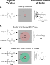
Figure 1
Figure 1
Basic stimulus. Physical variation is shown in the left column and perceived variation at the center is shown in the right column. (a) A circle within a dark gap was sinusoidally modulated along an L/(L + M) direction of MacLeod–Boynton color space. The perceived variation at the center is shown on the right. The difference between the perceived peak and trough of the variation is the perceived modulation depth. (b) The center and surround (separated by a dark gap) were temporally modulated at the same temporal frequency and had the same phase. The perceived variation at the center (shown at right) was reduced relative to (a). (c) The center and surround (separated by a dark gap) were temporally modulated at the same temporal frequency but in counterphase. The perceived variation at the center (shown at right) was enhanced relative to (a).
Figure 2
Figure 2
The experimental setup. (a) Dichoptic presentation. The test stimulus was presented on the top half of the monitor and the matching stimulus presented on the bottom half. The left and right sides of the monitor were presented to the left and right eyes, respectively, using a haploscope. In dichoptic presentation, the temporally modulating surround was presented to one eye and the temporally modulating center to the other eye. (b) Monocular presentation. Both the temporally modulating center and surround were presented to the same eye. (c) The fused percept.
Figure 3
Figure 3
Measurements with the surround modulation at 0.025 Michelson contrast. Each column shows results for one observer. The three rows show measurements for the three temporal frequencies. In each panel, the perceived modulation depth is plotted on the vertical axis, and the center–surround phase difference is on the horizontal axis. Monocular results are shown by red circles and dichoptic results by green circles. The blue square shows the perceived modulation depth with a steady surround. Lines through the plotted points are fits of the model in Equation 1 (see text). Error bars indicate one standard error of the mean.
Figure 4
Figure 4
As Figure 3, except with surround modulation at 0.05 Michelson contrast.
Figure 5
Figure 5
Parameter estimates from the model in Equation 1. Each column shows values for one observer. The two rows correspond to values for surround strength and neural phase difference, respectively. In the top row, surround strength RS is on the vertical axis and temporal frequency is on the horizontal axis. Monocular (dichoptic) surround strengths are shown by red (green) circles, with solid and dashed lines corresponding to the 0.025 and 0.05 Michelson contrast surrounds, respectively. In the bottom row, phase shift φ is on the vertical axis and temporal frequency is on the horizontal axis. Symbol coding is the same as for the panels in the top row. Surround strengths and phases were estimated using Equation 2. The standard errors for each parameter estimate (shown by the vertical bars) were calculated using a bootstrapping method as described in the text.
Figure 6
Figure 6
Estimates of isolated monocular and binocular surround strengths (see text and Equation 3), for all three observers. Surround strength is shown on the vertical axis and temporal frequency is on the horizontal axis. Estimates of isolated monocular (binocular) surround strength are shown by the red (green) circles, with the solid and dashed lines corresponding to the 0.025 and 0.05 Michelson contrast surrounds, respectively. Standard errors were again calculated using a bootstrapping technique.

Similar articles

References

    1. Autrusseau F., Shevell S. K. (2006). Temporal nulling of induction from spatial patterns modulated in time. Visual Neuroscience , 23 (3-4), 479–482 - PubMed
    1. Barbur J. L., Spang K. (2008). Colour constancy and conscious perception of changes of illuminant. Neuropscyhologia , 46, 853–863 - PubMed
    1. Christiansen J. H., D'Antona A. D., Shevell S. K. (2009). The neural pathways mediating color shifts induced by temporally varying light. Journal of Vision , 9 (5): 13 1–10, http://www.journalofvision.org/content/9/5/26, doi:10.1167/9.5.26. [PubMed] [Article] - PMC - PubMed
    1. Conway B. R. (2001). Spatial structure of cone inputs to color cells in alert macaque primary visual cortex (V-1). Journal of Neuroscience , 21 (8), 2768–2783 - PMC - PubMed
    1. Conway B. R., Hubel D. H., Livingstone M. (2002). Color contrast in macaque V1. Cerebral Cortex , 12, 915–925 - PubMed

Publication types

LinkOut - more resources