Alternative technique of cervical spinal stabilization employing lateral mass plate and screw and intra-articular spacer fixation
- PMID: 24744562
- PMCID: PMC3980556
- DOI: 10.4103/0974-8237.128527
Alternative technique of cervical spinal stabilization employing lateral mass plate and screw and intra-articular spacer fixation
Abstract
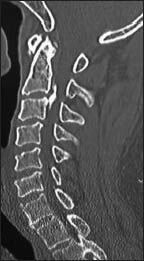
Aim: The author discusses an alternative technique of segmental cervical spinal fixation.
Material and methods: The subtleties of the technique are discussed on the basis of experience with 3 cases with a follow-up of between 30 and 36 months.
Technique: The technique involves debridement of facetal articular cartilage, distraction of facets, jamming of 'Goel spacer' into the articular cavity and fortification of the fixation by lateral mass plate and screw fixation. The 'double-insurance' method of fixation is safe for vertebral artery, nerve roots and spinal neural structures and the fixation is strong.
Conclusions: The discussed technique is safe and provides a strong fixation and a ground for ultimate arthrodesis.
Keywords: Goel spacer; intraarticular; plate; screw; transarticular.
Conflict of interest statement
Figures


Similar articles
-
Quantitative morphometric analysis of the lumbar vertebral facets and evaluation of feasibility of lumbar spinal nerve root and spinal canal decompression using the Goel intraarticular facetal spacer distraction technique: A lumbar/cervical facet comparison.J Craniovertebr Junction Spine. 2014 Oct;5(4):157-62. doi: 10.4103/0974-8237.147079. J Craniovertebr Junction Spine. 2014. PMID: 25558146 Free PMC article.
-
Only fixation for cervical spondylosis: Report of early results with a preliminary experience with 6 cases.J Craniovertebr Junction Spine. 2013 Jul;4(2):64-8. doi: 10.4103/0974-8237.128531. J Craniovertebr Junction Spine. 2013. PMID: 24744564 Free PMC article.
-
Caudally Directed Inferior Facetal and Transfacetal Screws for C1-C2 and C1-2-3 Fixation.World Neurosurg. 2017 Apr;100:236-243. doi: 10.1016/j.wneu.2017.01.015. Epub 2017 Jan 16. World Neurosurg. 2017. PMID: 28093348
-
Facetal distraction as treatment for single- and multilevel cervical spondylotic radiculopathy and myelopathy: a preliminary report.J Neurosurg Spine. 2011 Jun;14(6):689-96. doi: 10.3171/2011.2.SPINE10601. Epub 2011 Mar 18. J Neurosurg Spine. 2011. PMID: 21417697
-
Only spinal fixation as treatment of prolapsed cervical intervertebral disc in patients presenting with myelopathy.J Craniovertebr Junction Spine. 2017 Oct-Dec;8(4):305-310. doi: 10.4103/jcvjs.JCVJS_137_17. J Craniovertebr Junction Spine. 2017. PMID: 29403240 Free PMC article.
Cited by
-
Artificial atlantoaxial and subaxial facetal joint - Proposal of models.J Craniovertebr Junction Spine. 2022 Apr-Jun;13(2):107-109. doi: 10.4103/jcvjs.jcvjs_74_22. Epub 2022 Jun 13. J Craniovertebr Junction Spine. 2022. PMID: 35837423 Free PMC article. No abstract available.
-
Can decompressive laminectomy for degenerative spondylotic lumbar and cervical canal stenosis become historical?J Craniovertebr Junction Spine. 2015 Oct-Dec;6(4):144-6. doi: 10.4103/0974-8237.167851. J Craniovertebr Junction Spine. 2015. PMID: 26692688 Free PMC article. No abstract available.
-
"Only fixation:" Simple act, but mammoth stride toward great aspiration in managing cervical spondylotic myelopathy.J Craniovertebr Junction Spine. 2015 Jul-Sep;6(3):137-9. doi: 10.4103/0974-8237.161597. J Craniovertebr Junction Spine. 2015. PMID: 26288551 Free PMC article. No abstract available.
-
When is inclusion of C2 vertebra in the fixation construct necessary in cases with multi-level spinal degeneration?J Craniovertebr Junction Spine. 2020 Oct-Dec;11(4):249-251. doi: 10.4103/jcvjs.JCVJS_167_20. Epub 2020 Nov 26. J Craniovertebr Junction Spine. 2020. PMID: 33824552 Free PMC article. No abstract available.
-
Atlantoaxial and subaxial cervical spinal fixation: Can it revolutionize surgical treatment of cervical myelopathy related to Ossified posterior longitudinal ligament?J Craniovertebr Junction Spine. 2017 Jan-Mar;8(1):5-8. doi: 10.4103/0974-8237.199876. J Craniovertebr Junction Spine. 2017. PMID: 28250630 Free PMC article. No abstract available.
References
-
- Goel A, Shah A. Facetal distraction as treatment for single- and multilevel cervical spondylotic radiculopathy and myelopathy: A preliminary report. J Neurosurg Spine. 2011;14:689–96. - PubMed
-
- Goel A, Shah A, Jadhav M, Nama S. Distraction of facets with intraarticular spacers as treatment for lumbar canal stenosis: Report on a preliminary experience with 21 cases. J Neurosurg Spine. 2013;19:672–7. - PubMed
-
- Roy-Camille R, Saillant G. Surgery of the cervical spine. 2. Dislocation. Fracture of the articular processes. Nouv Presse Med. 1972;1:2484–5. - PubMed
LinkOut - more resources
Full Text Sources
Other Literature Sources