Review
. 2014 Apr 14;20(14):3719-37.
doi: 10.3748/wjg.v20.i14.3719.

Natural history of hepatic metastases from colorectal cancer--pathobiological pathways with clinical significance

Affiliations
Review

Natural history of hepatic metastases from colorectal cancer--pathobiological pathways with clinical significance

Konstantinos A Paschos et al. World J Gastroenterol. .

Abstract

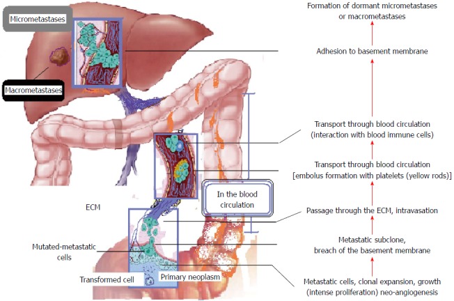
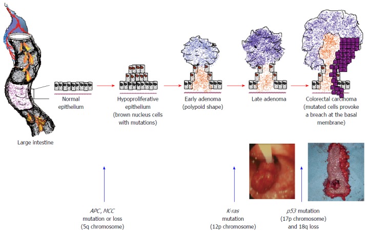
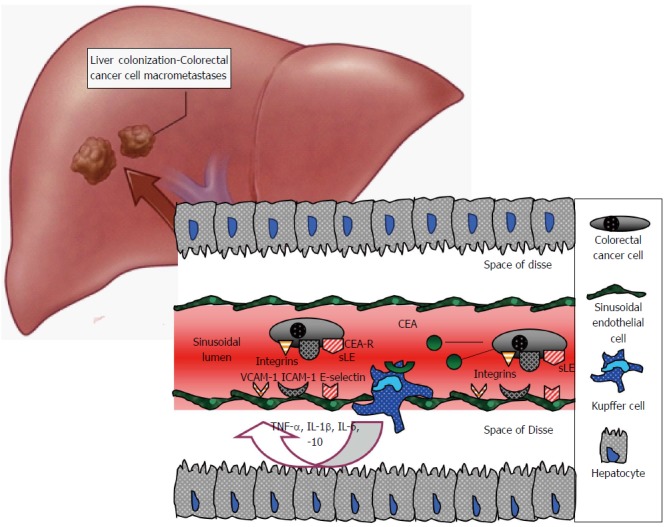
Colorectal cancer hepatic metastases represent the final stage of a multi-step biological process. This process starts with a series of mutations in colonic epithelial cells, continues with their detachment from the large intestine, dissemination through the blood and/or lymphatic circulation, attachment to the hepatic sinusoids and interactions with the sinusoidal cells, such as sinusoidal endothelial cells, Kupffer cells, stellate cells and pit cells. The metastatic sequence terminates with colorectal cancer cell invasion, adaptation and colonisation of the hepatic parenchyma. All these events, termed the colorectal cancer invasion-metastasis cascade, include multiple molecular pathways, intercellular interactions and expression of a plethora of chemokines and growth factors, and adhesion molecules, such as the selectins, the integrins or the cadherins, as well as enzymes including matrix metalloproteinases. This review aims to present recent advances that provide insights into these cell-biological events and emphasizes those that may be amenable to therapeutic targeting.

Keywords: Colorectal cancer; Liver metastasis; Liver sinusoids; Metastatic cascade; Neovascularization.


Figures

Figure 1
Figure 1
The invasion-metastasis cascade[1,178]. Successive interrelated stages/steps until foreign tissue colonisation and the formation of macrometastases. The paradigm of colorectal liver metastasis, from the large intestine through the superior and inferior mesenteric veins and the portal vein to the liver. ECM: Extra cellular matrix.
Figure 2
Figure 2
A sequence of mutations for colorectal epithelial cells generates the formation of polypoids, adenomas and finally carcinoma[15,16,19]. Polypoid and carcinoma clinical photos (authors’ archives) underneath the figures.
Figure 3
Figure 3
Various cellular types[22,23,112]. Various cellular types (resident: fibroblasts, endothelial cells and neurons, or recruited: macrophages, neutrophils and lymphocytes) which mediate cancer progression and growth in the colorectal microenvironment. bFGF: Basic fibroblast growth factor; CAF: Cancer associated fibroblasts; ECM: Extracellular matrix; EGF: Epidermal growth factor; EMT: Epithelial-to-mesenchymal transition; HGF: Hepatocyte growth factor; IDO: Indoleamine 2,3-dioxygenase; IGF: Insulin growth factor; IL-10: Interleukin 10; MMP: Matrix metalloprotease; NO: Nitric oxide; OPN: Osteopontin; PDGF-β: Platelet-derived growth factor-beta; PGE2: Prostaglandin E2; SDF-1: Stromal cell-derived factor-1; TAM: Tumor-associated macrophages; TGF-β: Transforming growth factor-beta; TNF-α: Tumour necrosis factor-alpha; VEGF: Vascular endothelial growth factor.
Figure 4
Figure 4
Sinusoidal endothelial cells. Fluorescence labeled (AcLDL) (A) and light microscopy (B). The cells have a cobblestone architecture on the sixth day in culture (authors’ archive). Magnification × 200. Scale bars = 100 μm. AcLDL: Acetylated low density lipoprotein.
Figure 5
Figure 5
The role of Kupffer cells in colorectal cell adhesion-arrest within the sinusoids during haematogenous liver metastases. Kupffer cells are activated through carcinoembryonic antigen (CEA), release multiple chemokines and stimulate sinusoidal endothelial cells to express adhesion molecules, inducing colorectal cancer cell arrest. CEA-R: CEA receptor; ICAM-1: Intercellular adhesion molecule 1; IL: Interleukin; sLE: Sialyl Lewis antigen; TNF-α: Tumour necrosis factor alpha; VCAM-1: Vascular cell adhesion molecule 1.
Figure 6
Figure 6
Hepatic stellate cells with their cytoplasmic processes, after 4 d in culture (authors’ archive). Light microscopy (magnification × 200). Scale bar = 100 μm.

References

    1. Valastyan S, Weinberg RA. Tumor metastasis: molecular insights and evolving paradigms. Cell. 2011;147:275–292. - PMC - PubMed
    1. Ghobrial IM. Myeloma as a model for the process of metastasis: implications for therapy. Blood. 2012;120:20–30. - PMC - PubMed
    1. Kumar S, Weaver VM. Mechanics, malignancy, and metastasis: the force journey of a tumor cell. Cancer Metastasis Rev. 2009;28:113–127. - PMC - PubMed
    1. Weinberg R. The biology of cancer. 1st ed. Taylor & Francis Group: Garland Science; 2007.
    1. Rejniak KA. Investigating dynamical deformations of tumor cells in circulation: predictions from a theoretical model. Front Oncol. 2012;2:111. - PMC - PubMed