. 2013 Dec 8;1(7):e00165.
doi: 10.1002/phy2.165. eCollection 2013 Dec 1.

Peptide transporter isoforms are discriminated by the fluorophore-conjugated dipeptides β-Ala- and d-Ala-Lys-N-7-amino-4-methylcoumarin-3-acetic acid

Affiliations

Peptide transporter isoforms are discriminated by the fluorophore-conjugated dipeptides β-Ala- and d-Ala-Lys-N-7-amino-4-methylcoumarin-3-acetic acid

Gabor Kottra et al. Physiol Rep. .

Abstract

Peptide transporters of the SLC15 family are classified by structure and function into PEPT1 (low-affinity/high-capacity) and PEPT2 (high-affinity/low-capacity) isoforms. Despite the differences in kinetics, both transporter isoforms are reckoned to transport essentially all possible di- and tripeptides. We here report that the fluorophore-conjugated dipeptide derivatives β-Ala-Lys-N-7-amino-4-methylcoumarin-3-acetic acid (β-AK-AMCA) and d-Ala-Lys-N-7-amino-4-methylcoumarin-3-acetic acid (d-AK-AMCA) are transported by distinct PEPT isoforms in a species-specific manner. Transport of the fluorophore peptides was studied (1) in vitro after heterologous expression in Xenopus oocytes of PEPT1 and PEPT2 isoforms from different vertebrate species and of PEPT1 and PEPT2 transporters from Caenorhabditis elegans by using electrophysiological and fluorescence methods and (2) in vivo in C. elegans by using fluorescence methods. Our results indicate that both substrates are transported by the vertebrate "renal-type" and the C. elegans "intestinal-type" peptide transporter only. A systematic analysis among species finds four predicted amino acid residues along the sequence that may account for the substrate uptake differences observed between the vertebrate PEPT1/nematode PEPT2 and the vertebrate PEPT2/nematode PEPT1 subtype. This selectivity on basis of isoforms and species may be helpful in better defining the structure-function determinants of the proteins of the SLC15 family.

Keywords: Caenorhabditis elegans; PEPT1; PEPT2; di‐ and tripeptide; substrate specificity; transporter.


Figures
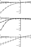
Figure 1.
Figure 1.
Current–voltage (I/V) relations induced by 1 mmol/L GQ (open circles) and by 1 mmol/L β‐AK‐AMCA (filled squares) in oocytes expressing (A) Caenorhabditis elegans PEPT1 (cePEPT1), (B) rabbit PEPT2 (rPEPT2), or (C) rabbit PEPT1 (rPEPT1). Please note the low, but significant transport current induced by β‐AK‐AMCA in cePEPT1, the higher current in rPEPT2 and the absence of β‐AK‐AMCA‐induced current in rPEPT1. Similar I/V relations have also been recorded in the other transporters listed in Table 1 (not shown).
Figure 2.
Figure 2.
Fluorescence (arbitrary units) induced by 3 h incubation in solutions containing 1 mmol/L β‐AK‐AMCA without or with 20 mmol/L GQ in oocytes expressing Caenorhabditis elegans (ce), human (h), rabbit (r), zebrafish (zf), mouse (m) PEPT1 and PEPT2 transporters, and rabbit chimeric transporters. NI, non‐injected. Dark columns: without β‐AK‐AMCA; shaded columns: with 1 mmol/L β‐AK‐AMCA; open columns: with 1 mmol/L β‐AK‐AMCA + 20 mmol/L GQ. For details see Materials and Methods.
Figure 3.
Figure 3.
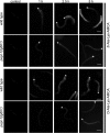
Time‐dependent uptake of β‐AK‐AMCA and d‐AK‐AMCA in wild type and peptide transporter deficient pept‐1(lg601) Caenorhabditis elegans. The animals were incubated for 10 min, 1 h, 2.5 h, and 5 h in medium containing 1 mmol/L β‐ or d‐AK‐AMCA and representative images are shown. The fluorescent signal is mostly detectable in the intestinal epithelial cells. The anterior end of the nematodes in the representative images is positioned to the top and the asterix mark the terminal bulb of the pharynx (*). Scale bars indicate 50 μm.
Figure 4.
Figure 4.
Molecular phylogenetic analysis performed on vertebrate PEPT1 and PEPT2 and nematode PEPT1 and PEPT2 by maximum likelihood (ML) method. The evolutionary history was inferred by using the ML method based on the Whelan and Goldman (2001) model. The tree with the highest log likelihood (−37,196.4871) is shown. Initial tree(s) for the heuristic search were obtained automatically as follows. When the number of common sites was <100 or less than one‐fourth of the total number of sites, the maximum parsimony method was used; otherwise BIONJ method with MCL distance matrix was used. A discrete Gamma distribution was used to model evolutionary rate differences among sites (five categories; +G, parameter = 0.9600). The rate variation model allowed for some sites to be evolutionarily invariable ([+I], 9.1163% sites). The tree is drawn to scale, with branch lengths measured in the number of substitutions per site. The analysis involved 114 amino acid sequences. All positions with less than 95% site coverage were eliminated. That is, fewer than 5% alignment gaps, missing data, and ambiguous bases were allowed at any position. There were a total of 630 positions in the final data set. Evolutionary analyses were conducted in MEGA5 (Tamura et al. 2011).
Figure 5.
Figure 5.
Sequence comparison performed on rabbit, human, mouse and zebrafish PEPT1, rabbit, human and zebrafish PEPT2, Caenorhabditis elegans PEPT1, and C. elegans PEPT2 amino acid sequences. Multiple sequence alignment was obtained by Clustal Omega (1.2.0) using default parameters. Consensus is reported. Candidate amino acid substitutions along the sequence are indicated with an arrow highlighted in red.

Similar articles

Cited by

References

    1. Abe H., Satoh M., Miyauchi S., Shuto S., Matsuda A., Kamo N. 1999. Conjugation of dipeptide to fluorescent dyes enhances its affinity for a dipeptide transporter (PEPT1) in human intestinal Caco‐2 cells. Bioconjug. Chem.; 10:24-31 - PubMed
    1. Allman E., Johnson D., Nehrke K. 2009. Loss of the apical V‐ATPase a‐subunit VHA‐6 prevents acidification of the intestinal lumen during a rhythmic behavior in C. elegans. Am. J. Physiol. Cell Physiol.; 297:C1071-C1081 - PMC - PubMed
    1. Bednarczyk D., Mash E. A., Aavula B. R., Wright S. H. 2000. NBD‐TMA: a novel fluorescent substrate of the peritubular organic cation transporter of renal proximal tubules. Pflugers Arch.; 440:184-192 - PubMed
    1. Biegel A., Gebauer S., Hartrodt B., Brandsch M., Neubert K., Thondorf I. 2005. Three‐dimensional quantitative structure‐activity relationship analyses of beta‐lactam antibiotics and tripeptides as substrates of the mammalian H+/peptide cotransporter PEPT1. J. Med. Chem.; 48:4410-4419 - PubMed
    1. Biegel A., Gebauer S., Brandsch M., Neubert K., Thondorf I. 2006. Structural requirements for the substrates of the H+/peptide cotransporter PEPT2 determined by three‐dimensional quantitative structure‐activity relationship analysis. J. Med. Chem.; 49:4286-4296 - PubMed

LinkOut - more resources