

Roles of the subfornical organ and area postrema in arterial pressure increases induced by 48-h water deprivation in normal rats
- PMID: 24744870
- PMCID: PMC3967674
- DOI: 10.1002/phy2.191
Roles of the subfornical organ and area postrema in arterial pressure increases induced by 48-h water deprivation in normal rats
Abstract
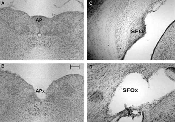
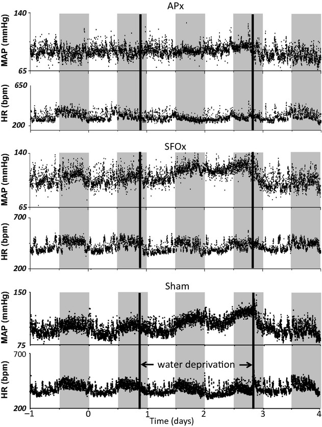
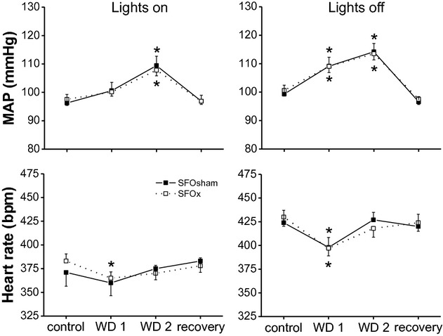
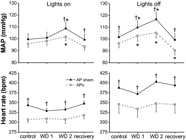
In rats, water deprivation (WD) increases arterial blood pressure (BP) in part due to actions of elevated osmolality in the brain to increase vasopressin levels and sympathetic activity. However, the osmoreceptors that mediate this response have not been identified. To test the hypothesis that osmoregulatory circumventricular organs are involved, BP and heart rate (HR) were continuously recorded telemetrically during 48 h of WD in normal rats with lesions (x) or sham lesions (sham) of the subfornical organ (SFO) or area postrema (AP). Although WD increased BP in SFOx and SFOsham rats, no significant difference in the hypertensive response was observed between groups. HR decreased transiently but similarly in SFOx and SFOsham rats during the first 24 h of WD. When water was reintroduced, BP and HR decreased rapidly and similarly in both groups. BP (during lights off) and HR were both lower in APx rats before WD compared to APsham. WD increased BP less in APx rats, and the transient bradycardia was eliminated. Upon reintroduction of drinking water, smaller falls in both BP and HR were observed in APx rats compared to APsham rats. WD increased plasma osmolality and vasopressin levels similarly in APx and APsham rats, and acute blockade of systemic V1 vasopressin receptors elicited similar depressor responses, suggesting that the attenuated BP response is not due to smaller increases in vasopressin or osmolality. In conclusion, the AP, but not the SFO, is required for the maximal hypertensive effect induced by WD in rats.
Keywords: Area postrema; blood pressure; heart rate; subfornical organ; water deprivation.
Figures
Similar articles
-
Osmolality: a physiological long-term regulator of lumbar sympathetic nerve activity and arterial pressure.Am J Physiol. 1999 Jun;276(6):R1579-86. doi: 10.1152/ajpregu.1999.276.6.R1579. Am J Physiol. 1999. PMID: 10362734
-
Water deprivation-induced sodium appetite and differential expression of encephalic c-Fos immunoreactivity in the spontaneously hypertensive rat.Am J Physiol Regul Integr Comp Physiol. 2010 May;298(5):R1298-309. doi: 10.1152/ajpregu.00359.2009. Epub 2010 Mar 3. Am J Physiol Regul Integr Comp Physiol. 2010. PMID: 20200133
-
Vasopressin acts in the subfornical organ to decrease blood pressure.Neuroendocrinology. 1997 Aug;66(2):130-5. doi: 10.1159/000127230. Neuroendocrinology. 1997. PMID: 9263210
-
Angiotensin and osmoreceptor inputs to the area postrema: role in long-term control of fluid homeostasis and arterial pressure.Clin Exp Pharmacol Physiol. 2000 May-Jun;27(5-6):443-9. doi: 10.1046/j.1440-1681.2000.03263.x. Clin Exp Pharmacol Physiol. 2000. PMID: 10831251 Review.
-
Vasopressin secretion: osmotic and hormonal regulation by the lamina terminalis.J Neuroendocrinol. 2004 Apr;16(4):340-7. doi: 10.1111/j.0953-8194.2004.01184.x. J Neuroendocrinol. 2004. PMID: 15089972 Review.
Cited by
-
Activation of the hypothalamic paraventricular nucleus by forebrain hypertonicity selectively increases tonic vasomotor sympathetic nerve activity.Am J Physiol Regul Integr Comp Physiol. 2015 Mar 1;308(5):R351-9. doi: 10.1152/ajpregu.00460.2014. Epub 2014 Dec 17. Am J Physiol Regul Integr Comp Physiol. 2015. PMID: 25519737 Free PMC article.
-
Hydration Status and Cardiovascular Function.Nutrients. 2019 Aug 11;11(8):1866. doi: 10.3390/nu11081866. Nutrients. 2019. PMID: 31405195 Free PMC article. Review.
References
-
- Anderson J. W., Washburn D. L., Ferguson A. V. 2000. Intrinsic osmosensitivity of subfornical organ neurons. Neuroscience; 100:539-547 - PubMed
-
- Anderson J. W., Smith P. M., Ferguson A. V. 2001. Subfornical organ neurons projecting to paraventricular nucleus: whole‐cell properties. Brain Res.; 921:78-85 - PubMed
-
- Blair M. L., Woolf P. D., Felten S. Y. 1997. Sympathetic activation cannot fully account for increased plasma renin levels during water deprivation. Am. J. Physiol.; 272:R1197-R1203 - PubMed
-
- Blessing W. W., Hedger S. C., Joh T. H., Willoughby J. O. 1987. Neurons in the area postrema are the only catecholamine‐ synthesizing cells in the medulla or pons with projections to the rostral ventrolateral medulla (C1‐area) in the rabbit. Brain Res.; 419:336-340 - PubMed
-
- Brooks V. L., Osborn J. W. 1995. Hormonal‐sympathetic interactions in long‐term regulation of arterial pressure: an hypothesis. Am. J. Physiol.; 268:R1343-R1358 - PubMed
Grants and funding
LinkOut - more resources
Full Text Sources
Other Literature Sources
Miscellaneous
