

doi: 10.1155/2014/524574.
Epub 2014 Mar 13.
A pilocytic astrocytoma mimicking a clinoidal meningioma
Affiliations
- PMID: 24744944
- PMCID: PMC3972933
- DOI: 10.1155/2014/524574
Item in Clipboard
A pilocytic astrocytoma mimicking a clinoidal meningioma
Case Rep Radiol.
2014.
Abstract
Pilocytic astrocytomas and meningiomas are benign, primary brain tumors that may involve the optic tract. Classically, the presence of a dural "tail" sign may differentiate a meningioma from other intracranial lesions. In this report, we describe a mass with the typical appearance of a clinoidal meningioma on magnetic resonance imaging (MRI) but postoperatively diagnosed as a pilocytic astrocytoma. This case illustrates the rare occurrence of a pilocytic astrocytoma mimicking a meningioma on MRI.
Figures
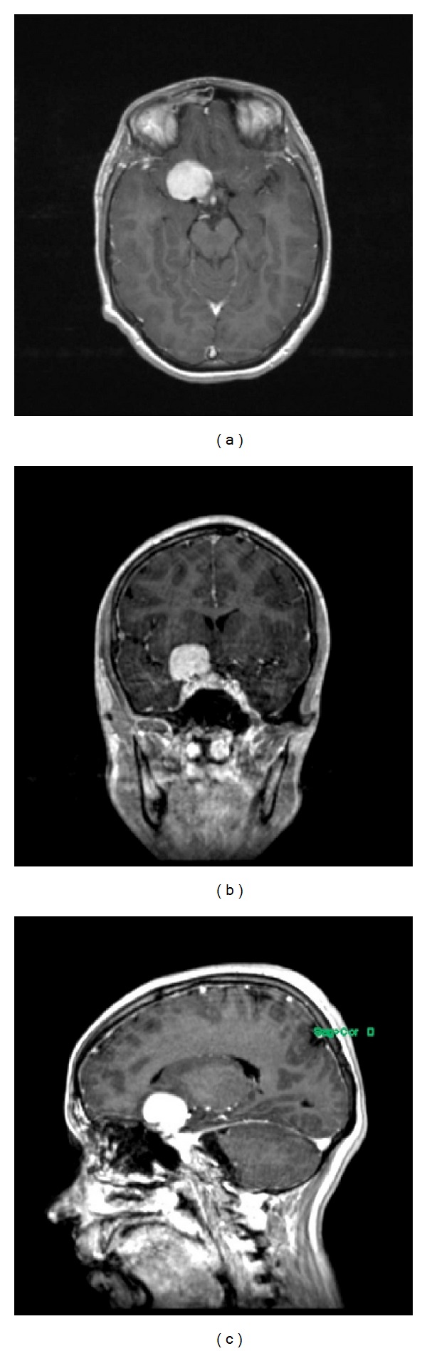
(a) Axial, (b) coronal, and (c) sagittal T1-weighted MRI showing a homogenously enhancing lesion, measuring 2.9 × 3.0 × 2.0 cm in the right clinoidal region. There is probable extension into the right optic canal, sphenoid sinus, right temporal fossa, and possibly right cavernous sinus, suggestive of a right clinoidal meningioma.
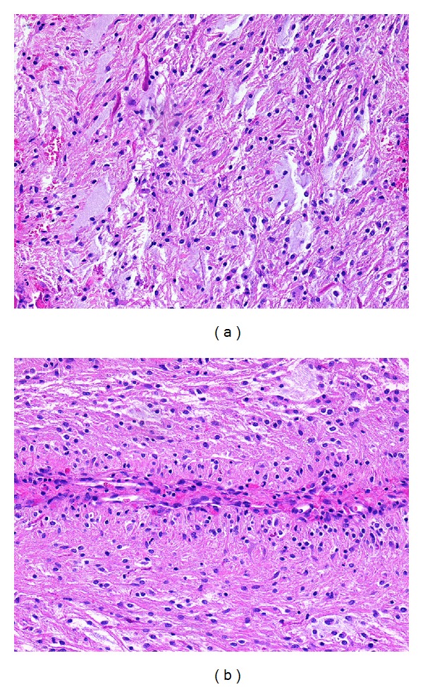
(a) The tumor consists of piloid cells and shows abundant mucin and Rosenthal fibers (upper left) (H&E stain; original magnification 200x). (b) A few perivascular pseudorosettes were noted, here in longitudinal section (H&E stain; original magnification 200x).
Similar articles
-
Supratentorial Pilocytic Astrocytoma Mimicking Convexity Meningioma with Early Anaplastic Transformation: A Case Report.Brain Tumor Res Treat. 2017 Oct;5(2):105-109. doi: 10.14791/btrt.2017.5.2.105. Epub 2017 Oct 31. Brain Tumor Res Treat. 2017. PMID: 29188212 Free PMC article.
-
Induction of gliosarcoma and atypical meningioma 13 years after radiotherapy of residual pilocytic astrocytoma in childhood.Pediatr Neurosurg. 2008;44(2):153-8. doi: 10.1159/000113120. Epub 2008 Jan 24. Pediatr Neurosurg. 2008. PMID: 18230932
-
Pilocytic astrocytomas: well-demarcated magnetic resonance appearance despite frequent infiltration histologically.Mayo Clin Proc. 1995 Aug;70(8):747-51. doi: 10.4065/70.8.747. Mayo Clin Proc. 1995. PMID: 7630212
-
Multiple cystic brain lesions in a patient with pilocytic astrocytoma.J Clin Neurosci. 2001 Jul;8(4):363-6. doi: 10.1054/jocn.2000.0802. J Clin Neurosci. 2001. PMID: 11437582 Review.
-
Holocord Pilocytic Astrocytoma in an Adult: A Rare Case Report and Review of the Literature.World Neurosurg. 2019 Jun;126:369-375. doi: 10.1016/j.wneu.2019.03.103. Epub 2019 Mar 19. World Neurosurg. 2019. PMID: 30902768 Review.
Cited by
-
Anterior Clinoid Metastasis as First Presentation of a Signet Ring Cell Carcinoma: An Intriguing Diagnosis.J Neurol Surg Rep. 2020 Jul;81(3):e46-e51. doi: 10.1055/s-0040-1712919. Epub 2020 Aug 14. J Neurol Surg Rep. 2020. PMID: 32818133 Free PMC article.
-
Supratentorial Pilocytic Astrocytoma Mimicking Convexity Meningioma with Early Anaplastic Transformation: A Case Report.Brain Tumor Res Treat. 2017 Oct;5(2):105-109. doi: 10.14791/btrt.2017.5.2.105. Epub 2017 Oct 31. Brain Tumor Res Treat. 2017. PMID: 29188212 Free PMC article.
-
Hypothalamic Hemangioma-like Pilocytic Astrocytoma in an Adult Patient: A Systematic Review with a Focus on Differential Diagnosis and Neurological Presentation.J Clin Med. 2024 Jun 17;13(12):3536. doi: 10.3390/jcm13123536. J Clin Med. 2024. PMID: 38930064 Free PMC article. Review.
-
Involvement of the Olfactory Apparatus by Gliomas.AJNR Am J Neuroradiol. 2020 Apr;41(4):712-717. doi: 10.3174/ajnr.A6471. Epub 2020 Mar 12. AJNR Am J Neuroradiol. 2020. PMID: 32165363 Free PMC article.
-
Anterior Clinoid Metastasis Removed Extradurally: First Case Report.J Neurol Surg Rep. 2018 Apr;79(2):e55-e62. doi: 10.1055/s-0038-1655773. Epub 2018 May 31. J Neurol Surg Rep. 2018. PMID: 29868330 Free PMC article.
References
-
- Listernick R, Charrow J, Greenwald M, Mets M. Natural history of optic pathway tumors in children with neurofibromatosis type 1: a longitudinal study. Journal of Pediatrics. 1994;125(1):63–66. - PubMed
-
- Qi ST, Lui Y, Pan J, et al. A radiopathological classification of dural tail sign of meningiomas. Journal of Neurosurgery. 2012;117(4):645–653. - PubMed
-
- Lusis EA, Scheithauer BW, Yachnis AT, et al. Meningiomas in pregnancy: a clinicopathological study of 17 cases. Neurosurgery. 2012;71(5):951–961. - PubMed
-
- Edwards JR, Kulwin CG, Martin SE, et al. Temporal and optic pathway pilomyxoid astrocytoma mimicking dural-based lesion: case report and review of the literature. Pediatric Neurosurgery. 2012;48(4):253–257. - PubMed
-
- Dirks PB, Jay V, Becker LE, et al. Development of anaplastic changes in low-grade astrocytomas of childhood. Neurosurgery. 1994;34(1):68–78. - PubMed
Grants and funding
LinkOut - more resources
Full Text Sources
Other Literature Sources
