. 2013 Oct 1;2(4):e26548.
doi: 10.4161/worm.26548.

C. elegans RNA-binding protein GLD-1 recognizes its multiple targets using sequence, context, and structural information to repress translation

Affiliations

C. elegans RNA-binding protein GLD-1 recognizes its multiple targets using sequence, context, and structural information to repress translation

Jung H Doh et al. Worm. .

Abstract

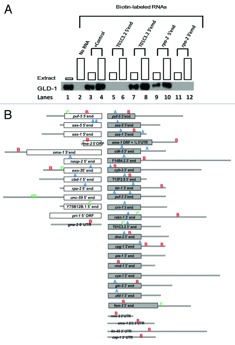
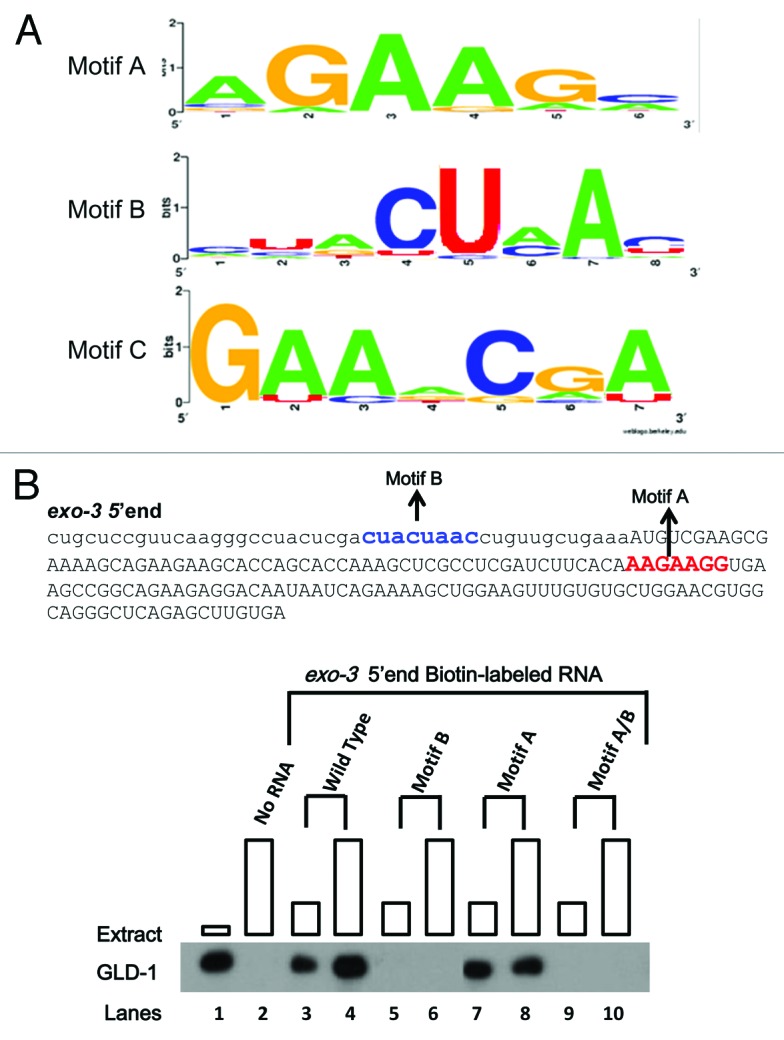
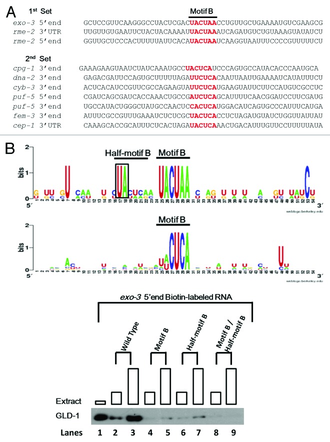
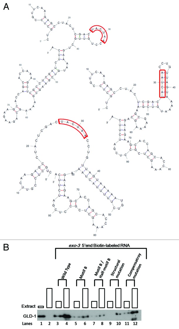
Caenorhabditis elegans GLD-1, a maxi-KH motif containing RNA-binding protein, has various functions mainly during female germ cell development, suggesting that it likely controls the expression of a selective group of maternal mRNAs. To gain an insight into how GLD-1 specifically recognizes these mRNA targets, we identified 38 biochemically proven GLD-1 binding regions from multiple mRNA targets that are among over 100 putative targets co-immunoprecipitated with GLD-1. The sequence information of these regions revealed three over-represented and phylogenetically conserved sequence motifs. We found that two of the motifs, one of which is novel, are important for GLD-1 binding in several GLD-1 binding regions but not in other regions. Further analyses indicate that the importance of one of the sequence motifs is dependent on two aspects: (1) surrounding sequence information, likely acting as an accessory feature for GLD-1 to efficiently select the sequence motif and (2) RNA secondary structural environment where the sequence motif resides, which likely provides "binding-site accessibility" for GLD-1 to effectively recognize its targets. Our data suggest some mRNAs recruit GLD-1 by a distinct mechanism, which involves more than one sequence motif that needs to be embedded in the correct context and structural environment.

Keywords: GLD-1; binding site accessibility; context dependency; mRNA targets; sequence motif.


Figures

None
Figure 1. GLD-1 binds to the 5′, 3′ or both ends of 32 mRNA targets. (A) GLD-1 binds to the 3′ end of T01C3.2 and the 5′end of MO4F3.1 RNAs. Western blot analysis of GLD-1/ Biotin-RNA pull-down assays of the 5′ and 3′ ends T01C3.2 and M04F3.1 mRNAs. All biotin-labeled RNAs (400 ng) were incubated with increasing amounts of wild-type cytosol extracts (50–150 ug) and then the RNA-protein complexes were isolated with Streptavidin-magnetic-beads. A western blot analysis was performed to detect the presence of GLD-1 in the complex. GLD-1 was probed in the cytosolic extract in the 1st lane, and no RNA was added to the binding reaction in the second lane. tra-2 3′UTR was used as a positive (+) control with increasing amount of cytosolic extract in the third and fourth lanes. Subsequent lanes represent binding reactions with 5′ and 3′ ends of T01C3.2 and M04F3.1 biotin-labeled RNAs. Boxes at each lane are proportional to the amount of extract used. (B) GLD-1 binds to 38 regions in 32 mRNA targets. 32 GLD-1 targets identified in the RIP-chip analysis were employed to identify GLD-1 binding regions. GLD-1 was able to bind to the 5′, 3′, or both ends of all targets. For each region, the GLD-1/biotin RNA binding assays and western blot analyses were performed at least twice to validate the consistency of the results. White and gray bars indicate the 5′ and 3′ coding regions, respectively, and the gray lines indicate the untranslated regions (UTRs). A Patser scan was performed to locate the over-represented motif A, B, and C identified through PhyloCon and PhyloNet within the 38 GLD-1 binding regions, which are marked at approximate locations. Blue “A,” red “B,” and green “C” represent the Motif A, Motif B, and Motif C, respectively.
None
Figure 2. Over-represented sequence Motifs A, B, and C were identified and examined for their importance in GLD-1 binding. (A) The sequences of 38 GBRs from 32 mRNA targets and their corresponding orthologous regions from four closely related nematodes were subjected to PhyloCon and PhyloNet analyses, and sequence Motif A and B (PhyloCon and PhyloNet) and C (PhyloNet) were identified as over-represented. Each motif is represented as a logo that shows a graphical representation of a nucleic acid multiple sequence alignment. The overall height of the stacked nucleotides within each motif indicates the degree of sequence conservation at the position, while the height of symbols within the stack indicates the relative frequency of each nucleic acid at the position. (B) Motif B is important for GLD-1 binding in R09B3.1 5′ end. Single or double mutations containing mutations in Motif B significantly reduces GLD-1 binding to the R09B3.1 3′ end. The RNA sequence of the R09B3.1 3′ end is shown (top). Sequence in lower-case represents the 5′UTR and in upper-case the first 150 nucleotides of the ORF. The Motif A and B are depicted in the sequence. The four most conserved nucleotides were mutated, where the Motif A was mutated to “AUCUUGG” while the Motif B is mutated “UAGAUU”. Biotin-RNA pull-down assays were performed with the wild-type and mutated R09B3.1 3′ end as described previously. Boxes at each lane are proportional to the amount of extract used.
None
Figure 3. The identification of the Half-motif B and its importance in GLD-1 binding. (A) The possibility that sequences surrounding Motif B may contribute in a context-dependent manner to GLD-1 binding was examined. Two sets of sequences were compiled. The first set contains three GBRs where mutations of the Motif B reduced or eliminated GLD-1 binding, indicating that the Motif Bs in these GBR are important for GLD-1 binding. The second set contains the seven GBRs where GLD-1 still binds well even though the Motif Bs are mutated, indicating that the Motif Bs in these regions have no biochemical importance. Next, these two data sets were aligned so that Motif B was centered within the alignment with additional ~25 nucleotides flanking either side of the Motif Bs. (B) With the hypothesis that adjacent sequences important for GLD-1 recognition should be conserved, two independent sequence alignments were performed to see if any nucleotides were conserved in the first but not in the second set. Sequence alignment matrixes of the two sets are shown. Motif B is highly conserved in both data sets. Also, a Uridine-Adenine (UA) site just upstream of Motif B is highly conserved in the sequence alignment of the first set, which is not detected in the sequence alignment of the second set. Sequences that immediately follow the conserved UA site in the first set represent a weakly conserved Motif B, (UA C/U U/A C/A G/A) and is therefore designated as a Half-motif B. (C) The importance of Half-motif B for GLD-1/RNA interaction was examined. A western blot of the GLD-1/ Biotin-RNA pull-down assay with the wild-type R09B3.1 5′end and corresponding mutations is shown. The first lane shows GLD-1 in cytosolic extract. Lanes 2 through 9 represent binding reactions with wild-type and mutated R09B3.1 5′end biotin-labeled RNAs (400 ng) with increasing amount of cytosolic extract (50 and 150 ug). The conserved “UA” dinucleotide of the Half-motif B was mutated to “AU” and the Motif B (“UACUAA”) was mutated “UAGAUU” within R09B3.1 5′end (see Fig. 3B). The mutation of the Half-motif B reduces GLD-1 binding as significantly as the mutation of the Motif B compared with the wild-type. A double mutation of both motifs appears to reduce GLD-1 binding further, where the residual GLD-1 bands are no longer detected. Boxes are each lane are proportional to the amount of extract used.
None
Figure 4. GLD-1 binding to the Motif B within R09B3.1 5′end is dependent on the RNA secondary structure. (A) The most dominant structures of exo-3 5′end of the wild-type, structural mutant, and compensatory mutant are shown as predicted by MFold. The Motif Bs within each structure are outlined in red. The Motif B in the wild-type R09B3.1 5′end predicted to be completely unstructured, likely rendering the Motif B highly accessible for GLD-1 binding. A structural mutation forces the Motif B to become structured while maintain the wild-type Motif B sequence. A compensatory mutation places the Motif B back into unstructured conformation, likely bringing back Motif B’s accessibility to GLD-1 (right). (B) Western blot analysis of biotin/RNA pull-down assays of wild-type R09B3.1 5′end and corresponding mutations. Binding reactions were performed as identical to the previous assays. GLD-1 was probed in the cytosolic extract in the first lane, and no RNA was subjected as a negative control in the second lane. GLD-1 binding is significantly reduced when the Motif B and/or Half-motif B are mutated within R09B3.1 5′end as shown previously. GLD-1 binding is also significantly reduced with the structural mutation of the R09B3.1 5′end, whereas the GLD-1 binding is rescued with the compensatory mutation, comparable to the wild-type.

Similar articles

Cited by

References

    1. Crittenden SL, Bernstein DS, Bachorik JL, Thompson BE, Gallegos M, Petcherski AG, Moulder G, Barstead R, Wickens M, Kimble J. A conserved RNA-binding protein controls germline stem cells in Caenorhabditis elegans. Nature. 2002;417:660–3. doi: 10.1038/nature754. - DOI - PubMed
    1. D’Agostino I, Merritt C, Chen PL, Seydoux G, Subramaniam K. Translational repression restricts expression of the C. elegans Nanos homolog NOS-2 to the embryonic germline. Dev Biol. 2006;292:244–52. doi: 10.1016/j.ydbio.2005.11.046. - DOI - PubMed
    1. Jones AR, Schedl T. Mutations in gld-1, a female germ cell-specific tumor suppressor gene in Caenorhabditis elegans, affect a conserved domain also found in Src-associated protein Sam68. Genes Dev. 1995;9:1491–504. doi: 10.1101/gad.9.12.1491. - DOI - PubMed
    1. Lublin AL, Evans TC. The RNA-binding proteins PUF-5, PUF-6, and PUF-7 reveal multiple systems for maternal mRNA regulation during C. elegans oogenesis. Dev Biol. 2007;303:635–49. doi: 10.1016/j.ydbio.2006.12.004. - DOI - PubMed
    1. Kuersten S, Goodwin EB. The power of the 3′ UTR: translational control and development. Nat Rev Genet. 2003;4:626–37. doi: 10.1038/nrg1125. - DOI - PubMed

LinkOut - more resources