Review
. 2014:2014:165897.
doi: 10.1155/2014/165897. Epub 2014 Mar 5.

MicroRNAs in the Neural Retina

Affiliations
Review

MicroRNAs in the Neural Retina

Kalina Andreeva et al. Int J Genomics. 2014.

Abstract

The health and function of the visual system rely on a collaborative interaction between diverse classes of molecular regulators. One of these classes consists of transcription factors, which are known to bind to DNA and control the transcription activities of their target genes. For a long time, it was thought that the transcription factors were the only regulators of gene expression. More recently, however, a novel class of regulators emerged. This class consists of a large number of small noncoding endogenous RNAs, namely, miRNAs. The miRNAs compose an essential component of posttranscriptional gene regulation, since they ultimately control the fate of gene transcripts. The retina, as a part of the central nervous system, is a well-established model for unraveling the molecular mechanisms underlying neuronal and glial functions. Numerous recent efforts have been made towards identification of miRNAs and their inferred roles in the visual pathway. In this review, we summarize the current state of our knowledge regarding the expression and function of miRNA in the neural retina and we discuss their potential uses as biomarkers for some retinal disorders.


Figures

Figure 1
Figure 1
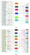
Two examples of mixed regulatory circuits, each containing a miRNA (oblate spheroid), linked to TFs (trapezoids) and coding gene targets (rectangles), all of which changed their expression in response to transient ischemia-reperfusion injury in the retina. The numbers and bold arrows represent fold change relative to sham control animals and up- /downregulation (resp.) of the miRNAs, TFs, and protein coding genes implicated in the mixed regulatory circuits. (a) Direct and indirect downregulation of two coding genes by the same miRNA (rno-miR-101a). At 24 h IR period, rno-miR-101a targets the protein coding genes Gria4 (glutamate receptor) and Nefl (neurofilament) directly. At the same time, rno-miR-101a can control these genes indirectly by targeting the TF Nptx1, which in turn targets Gria4 and Nefl. (b) miRNA targets several TFs which act together to coordinate the reduced expression of a single gene. At 24 h IR period, rno-miR-99a targets the protein coding gene Icam1 (adhesion molecule) directly. At the same time, rno-miR-99a can control this gene indirectly by targeting 3TFs (Arpc1b, Cebpb, and Stat3), which in turn target Icam1.
Figure 2
Figure 2
Regulatory subnetworks for two time points following transient retinal ischemia-reperfusion injury. The networks contain three types of molecules: miRNAs (shown in the left column), TFs (shown as trapezoids in the center), and coding gene targets (shown in the right column). All of these molecules changed their expression in response to IR injury twofold or more, relative to their sham controls. The arrows next to each gene correspond to gene regulation: uppointed arrows indicate increased gene expression; downpointed arrows indicate decreased gene expression. TFs, miRNAs, and coding genes that are linked with the same colored line indicate that the miRNAs target the TFs and at the same time, both of these regulators cotarget the coding genes. With the notable exception of Cebpb, there were no common target coding genes or TFs in the 2 subnetworks, even though they shared 19 common miRNAs. (a) Regulatory subnetwork consisting of miRNAs, TFs, and target genes, which changed their expression at the 24 h IR injury time point relative to their sham controls (FC ≥ 2). At this post-IR time point, six TFs were fine-tuned by 29 miRNAs to jointly regulate 17 target genes. (b) Regulatory subnetwork consisting of miRNAs, TFs, and target genes, which changed their expression at 7 days after IR injury relative to their sham controls (FC ≥ 2). At this post-IR time point, eight TFs were regulated by 30 miRNAs to jointly control the expression of 31 target genes.
Figure 3
Figure 3
The 3'UTR of the intronless Forkhead Box E1 gene (foxe1), located on chromosome 5 (X), is targeted by 10 miRNAs (rectangles with different colors). All the miRNAs are localized on different chromosomes in the rat genome (colored chromosome corresponds to miRNAs of the same color). The 10 miRNAs are upregulated in response to retinal IR-injury in rat retinal ischemic model while the expression level of foxe1 is reduced.

References

    1. Tarver JE, Donoghue PC, Peterson KJ. Do miRNAs have a deep evolutionary history? Bioessays. 2012;34(10):857–866. - PubMed
    1. Lim LP, Glasner ME, Yekta S, Burge CB, Bartel DP. Vertebrate microRNA genes. Science. 2003;299(5612):p. 1540. - PubMed
    1. Bandyra KJ, Said N, Pfeiffer V, Gorna MW, Vogel J, Luisi BF. The seed region of a small RNA drives the controlled destruction of the target mRNA by the endoribonuclease RNase E. Molecular Cell. 2012;47(6):943–953. - PMC - PubMed
    1. Huntzinger E, Izaurralde E. Gene silencing by microRNAs: contributions of translational repression and mRNA decay. Nature Reviews Genetics. 2011;12(2):99–110. - PubMed
    1. Davis BN, Hata A. Regulation of MicroRNA biogenesis: a miRiad of mechanisms. Cell Communication and Signaling. 2009;7(18):7–18. - PMC - PubMed

LinkOut - more resources