. 2014 Jun;13(6):951-62.
doi: 10.1039/c3pp50442e.

Photoconversion changes bilin chromophore conjugation and protein secondary structure in the violet/orange cyanobacteriochrome NpF2164g3' [corrected]

Affiliations

Photoconversion changes bilin chromophore conjugation and protein secondary structure in the violet/orange cyanobacteriochrome NpF2164g3' [corrected]

Sunghyuk Lim et al. Photochem Photobiol Sci. 2014 Jun.

Erratum in

  • Photochem Photobiol Sci. 2014 Sep;13(9):1360

Abstract

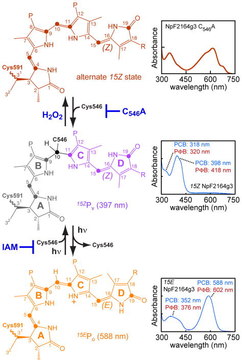
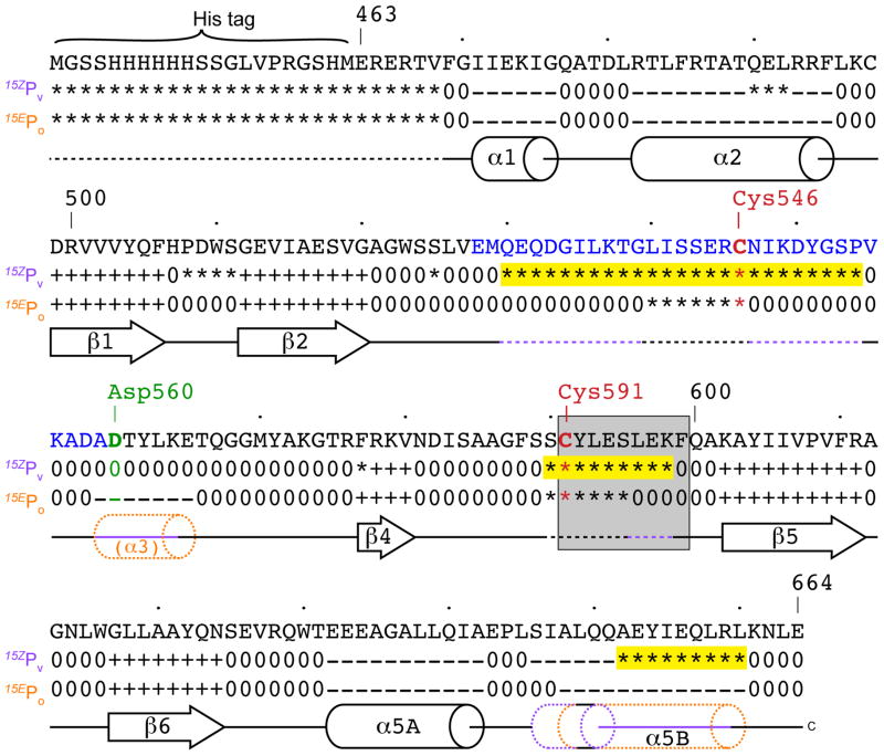
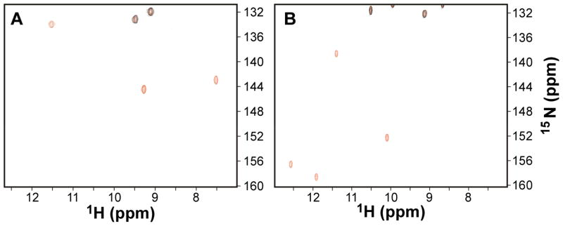
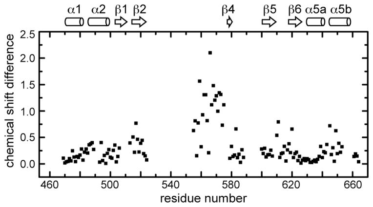
Cyanobacteriochromes (CBCRs) are cyanobacterial photoreceptors distantly related to phytochromes. All CBCRs examined to date utilize a conserved Cys residue to form a covalent thioether linkage to the bilin chromophore. In the insert-Cys CBCR subfamily, a second conserved Cys can covalently link to the bilin C10 methine bridge, allowing detection of near-UV to blue light. The best understood insert-Cys CBCR is the violet/orange CBCR NpF2164g3 from Nostoc punctiforme, which has a stable second linkage in the violet-absorbing dark state. Photoconversion of NpF2164g3 leads to elimination of the second linkage and formation of an orange-absorbing photoproduct. We recently reported NMR chemical shift assignments for the orange-absorbing photoproduct state of NpF2164g3. We here present equivalent information for its violet-absorbing dark state. In both photostates, NpF2164g3 is monomeric in solution and regions containing the two conserved Cys residues essential for photoconversion are structurally disordered. In contrast to blue light receptors such as phototropin, NpF2164g3 is less structurally ordered in the dark state than in the photoproduct. The insert-Cys insertion loop and C-terminal helix exhibit light-dependent structural changes. Moreover, a motif containing an Asp residue also found in other CBCRs and in phytochromes adopts a random-coil structure in the dark state but a stable α-helix structure in the photoproduct. NMR analysis of the chromophore is consistent with a less ordered dark state, with A-ring resonances only resolved in the photoproduct. The C10 atom of the bilin chromophore exhibits a drastic change in chemical shift upon photoconversion, changing from 34.5 ppm (methylene) in the dark state to 115 ppm (methine) in the light-activated state. Our results provide structural insight into the two-Cys photocycle of NpF2164g3 and the structurally diverse mechanisms used for light perception by the larger phytochrome superfamily.


Figures

Fig. 1
Fig. 1
Changes in CBCR secondary structure upon photoconversion. (A) Topology diagrams for the DXCF CBCR TePixJ, , based on solution structures of the blue-absorbing dark state (left) and green-absorbing photoproduct (right). (B) Topology diagrams for the insert-Cys CBCR NpF2164g3 as violet-absorbing dark state (left, this work) and orange-absorbing photoproduct (right). For NpF2164g3, the insert-Cys CBCR insertion loop (blue) is highlighted. For both proteins, critical Cys residues (red) are highlighted, regions not resolved in NMR secondary structure determination are dashed, and the α4 helix containing the Cys covalently linked to the bilin A-ring (Cys591 in NpF2164g3) is shaded grey where present.
Fig. 2
Fig. 2
The violet/orange photocycle of NpF2164g3. Bilin chromophore structures are shown on the left, absorption spectra are shown on the right, and experimental perturbations of the photocycle are highlighted in blue. IAM, iodoacetamide. The C18 sidechain varies with bilin (phycocyanobilin or PCB, R=Et; phytochromobilin or PΦB, R=Vn). A 15Z red-absorbing state (top) is observed experimentally upon mutation of Cys546 or upon treatment of the 15ZPV dark state with hydrogen peroxide. In wild-type NpF2164g3, the 15ZPV dark state is formed (middle). In the 15ZPV state, Cys546 is covalently attached to the bilin C10 atom. This splits the conjugated system of the bilin chromophore in two. The system formed by the C- and D-rings exhibits a red-shift with PΦB, while that formed by the A- and B-rings does not. Photoisomerization of the 15,16-double bond leads to elimination of the second linkage, restoring conjugation across the C10 methine bridge in the 15EPo state (bottom). Carbons, rings, and the configuration of the 15,16-double bond are indicated. Filled circles indicate carbon atoms labeled by bilin biosynthesis using 5-13C ALA. Grey, purple, and orange indicate conjugated systems absorbing at 322, 397, and 588 nm, respectively.
Fig. 3
Fig. 3
15N NMR relaxation data and protein backbone dynamics for individual photostates of NpF2164g3. (A) Light-activated state of NpF2164g3: 15N longitudinal relaxation rate constant, R1 (top panel), 15N transverse relaxation rate constant, R2 (middle panel), heteronuclear NOE (bottom panel) plotted as a function of residue number. (B) Dark-state of NpF2164g3: R1 (top panel) and R2 (bottom panel) are plotted as a function of residue number.
Fig. 4
Fig. 4
Secondary structure of NpF2164g3. Secondary structural elements were derived from analysis of chemical shift index and sequential NOE patterns. The chemical shift index sign (+, − or 0) is indicated underneath each residue. Residues in the insert-Cys insertion loop are colored blue. The two cysteine residues (Cys546 and Cys591) linked to the dark-state chromophore are colored red. The Asp-motif residue Asp560 is colored green. Unassigned residues are marked by an asterisk. Residues in the grey box are helical in AnPixJ (Fig. 2). Regions that are exchange broadened in the dark state are highlighted yellow. Regions of variable secondary structure are color-coded.
Fig. 5
Fig. 5
Two-dimensional 15N-1H HSQC NMR spectra of NpF2164g3 (pyrrole region). (A) NMR spectrum of 15N-labeled NpF2164g3 in the dark-adapted state containing a 14N-labeled bilin chromophore (black) overlaid onto that containing a 15N-labeled bilin chromophore (red). (B) NMR spectrum of light-activated NpF2164g3 containing a 14N-labeled bilin chromophore (black) overlaid onto that containing a 15N-labeled bilin chromophore (red). The non-pyrrole resonances (black at 15N ~ 132 ppm) are due to tryptophan indole side-chain resonances from the 15N-labeled protein. Pyrrole resonances are shown in red.
Fig. 6
Fig. 6
13C NMR spectra of NpF2164g3 with attached bilin chromophore containing 13C labeling (at C4, C5, C9, C10, C11, C15, and C19) in the dark-adapted state (A), light-activated state (B), and dark-minus-light difference spectrum (C).
Fig. 7
Fig. 7
Light-dependent structural changes in NpF2164g3 plotted as a function of residue number. Chemical shift differences between residues in the dark-state and light-activated states (CSD as defined in the text) are plotted.
Fig. 8
Fig. 8
Hypothetical progressive dimerization model for function of CBCRs in tandem. We consider a simplified CBCR photosensor, comprising an N-terminal violet/orange CBCR, a red/green CBCR, and a C-terminal output domain that requires dimerization for function. When both proteins are in the dark state (top left), the C-terminal α5 helix of each CBCR domain is unstable and the photoreceptor is largely monomeric. Photoconversion of the N-terminal CBCR domain by violet light (top right) results in formation of a stable helix connecting the two CBCR domains. Photoconversion of the C-terminal CBCR domain by red light (bottom left) results in formation of a stable helix connecting the red/green CBCR domain to the output domain. Photoconversion of both CBCR domains (bottom right) provides two stable helices and allows maximal formation of the stable, biologically active output-domain dimer. V, violet; R, red.

References

    1. Yeh KC, Wu SH, Murphy JT, Lagarias JC. A cyanobacterial phytochrome two-component light sensory system. Science. 1997;277:1505–1508. - PubMed
    1. Wagner JR, Brunzelle JS, Forest KT, Vierstra RD. A light-sensing knot revealed by the structure of the chromophore binding domain of phytochrome. Nature. 2005;438:325–331. - PubMed
    1. Wagner JR, Zhang J, Brunzelle JS, Vierstra RD, Forest KT. High resolution structure of Deinococcus bacteriophytochrome yields new insights into phytochrome architecture and evolution. J Biol Chem. 2007;282:12298–12309. - PubMed
    1. Yang X, Stojkovic EA, Kuk J, Moffat K. Crystal structure of the chromophore binding domain of an unusual bacteriophytochrome, RpBphP3, reveals residues that modulate photoconversion. Proc Natl Acad Sci USA. 2007;104:12571–12576. - PMC - PubMed
    1. Essen LO, Mailliet J, Hughes J. The structure of a complete phytochrome sensory module in the Pr ground state. Proc Natl Acad Sci USA. 2008;105:14709–14714. - PMC - PubMed

Publication types