Review
. 2014:73:175-204.
doi: 10.1016/B978-0-12-800223-0.00004-9.

Biology of the major facilitative folate transporters SLC19A1 and SLC46A1

Affiliations
Review

Biology of the major facilitative folate transporters SLC19A1 and SLC46A1

Zhanjun Hou et al. Curr Top Membr. 2014.

Abstract

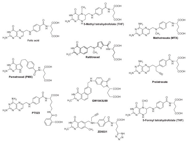
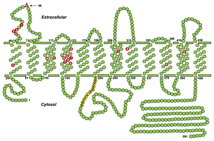
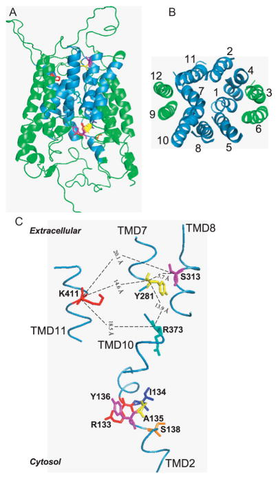
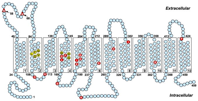
This chapter focuses on the biology of the major facilitative membrane folate transporters, the reduced folate carrier (RFC), and the proton-coupled folate transporter (PCFT). Folates are essential vitamins, and folate deficiency contributes to a variety of heath disorders. RFC is ubiquitously expressed and is the major folate transporter in mammalian cells and tissues. PCFT mediates intestinal absorption of dietary folates. Clinically relevant antifolates such as methotrexate (MTX) are transported by RFC, and the loss of RFC transport is an important mechanism of MTX resistance. PCFT is abundantly expressed in human tumors and is active under pH conditions associated with the tumor microenvironment. Pemetrexed (PMX) is an excellent substrate for PCFT as well as for RFC. Novel tumor-targeted antifolates related to PMX with selective membrane transport by PCFT over RFC are being developed. The molecular picture of RFC and PCFT continues to evolve relating to membrane topology, N-glycosylation, energetics, and identification of structurally and functionally important domains and amino acids. The molecular bases for MTX resistance associated with loss of RFC function, and for the rare autosomal recessive condition, hereditary folate malabsorption (HFM), attributable to mutant PCFT, have been established. From structural homologies to the bacterial transporters GlpT and LacY, homology models were developed for RFC and PCFT, enabling new mechanistic insights and experimentally testable hypotheses. RFC and PCFT exist as homo-oligomers, and evidence suggests that homo-oligomerization of RFC and PCFT monomeric proteins may be important for intracellular trafficking and/or transport function. Better understanding of the structure and function of RFC and PCFT should facilitate the rational development of new therapeutic strategies for cancer as well as for HFM.

Keywords: Antifolate; Folate; Oligomerization; Proton-coupled folate transporter; Reduced folate carrier; Transporter.


Figures

Figure 4.1
Figure 4.1
Structures of folates and clinically relevant antifolates. RFC substrates including MTX, PMX, ratitrexed, pralatrexate, 5-methyl THF, and 5-formyl THF are all excellent PCFT substrates. However, the antifolates PT523 and GW1843U89 do not appear to be transported by PCFT. RFC has a low affinity but PCFT has a high affinity for folic acid. GW1843U89 is an excellent substrate for human RFC, but a poor substrate for murine RFC.
Figure 4.2
Figure 4.2
Human RFC topology model. A topology model is shown for human RFC, with 12 TMDs, internal N- and C-termini, and a loop domain connecting TMD6 and 7. The structurally and functionally important amino acids, as described in the text, are shown as red circles. A conserved stretch of amino acids (Lys204–Arg214) in the TMD6/TMD7 loop domain, which is important for transport activity, is shown as yellow circles. N-glycosylation occurs at Asn58, which is labeled as a red triangle.
Figure 4.3
Figure 4.3
Three-dimensional (3D) homology models of human RFC. A 3D model for human RFC is presented, based on structure alignments between RFC and LacY/GlpT and experimental data. (A) A side view of the RFC for which the extended C-terminal segment is truncated at Lys479. TMD1, TMD2, TMD4, and TMD5 of the N-terminal region and TMD7, TMD8, TMD10, and TMD11 of the C-terminal region are involved in formation of the hydrophilic binding site for anionic folates (colored sky blue). TMD3, TMD6, TMD9, and TMD12 are buried in the lipid bilayer and do not directly participate in substrate binding (colored green). Panel (A) also depicts key amino acids (shown in assorted colors) that may contribute to the binding pocket for anionic folate substrates, as described in the text. (B) A cytosolic view of only the TMD segments of the human RFC molecule so that the order of helix packing can be seen easily. TMD coloring is the same as described in (A). (C) Enhanced view of the hypothetical substrate-binding site comprising the same key amino acids depicted in (A), including Lys411, Ser313, Tyr281, and Arg373, as described in the text. Other residues that may contribute to the substrate-binding pocket are also shown and include Arg133, Ile134, Ala135, Tyr136, and Ser138. The physical distances between the αcarboxyl groups of Lys411, Ser313, Tyr281, and Arg373 are given in angstroms. This figure was originally published in The Journal of Biological Chemistry, by Hou et al. (2006). © The American Society for Biochemistry and Molecular Biology.
Figure 4.4
Figure 4.4
Schematic structure of human PCFT membrane topology. A topology model is shown for human PCFT, with 12 TMDs and internal N- and C-termini. Structurally or functionally important amino acids, as determined from published mutagenesis studies and in patients with hereditary folate malabsorption (HFM), are shown as red circles. GXXXG putative oligomerization motifs are shown as yellow circles (Phe157 and Gly158 in the G155XXXG159 motif are shown as red circles because they are also structurally and functionally important). N-glycosylation occurs at Asn58 and Asn68 (shown as red triangles).

Similar articles

Cited by

References

    1. Abramson J, Smirnova I, Kasho V, Verner G, Kaback HR, Iwata S. Structure and mechanism of the lactose permease of Escherichia coli. Science. 2003;301(5633):610–615. - PubMed
    1. Assaraf YG, Babani S, Goldman ID. Increased activity of a novel low pH folate transporter associated with lipophilic antifolate resistance in Chinese hamster ovary cells. The Journal of Biological Chemistry. 1998;273(14):8106–8111. - PubMed
    1. Atabay B, Turker M, Ozer EA, Mahadeo K, Diop-Bove N, Goldman ID. Mutation of the proton-coupled folate transporter gene (PCFT-SLC46A1) in Turkish siblings with hereditary folate malabsorption. Pediatric Hematology and Oncology. 2010;27(8):614–619. - PMC - PubMed
    1. Borzutzky A, Crompton B, Bergmann AK, Giliani S, Baxi S, Martin M, et al. Reversible severe combined immunodeficiency phenotype secondary to a mutation of the proton-coupled folate transporter. Clinical Immunology. 2009;133(3):287–294. - PMC - PubMed
    1. Brigle KE, Spinella MJ, Sierra EE, Goldman ID. Characterization of a mutation in the reduced folate carrier in a transport defective L1210 murine leukemia cell line. The Journal of Biological Chemistry. 1995;270(39):22974–22979. - PubMed

Publication types

MeSH terms

Substances