Review
. 2014:209:91-129.
doi: 10.1016/B978-0-444-63274-6.00006-0.

The integrative role of the sigh in psychology, physiology, pathology, and neurobiology

Affiliations
Review

The integrative role of the sigh in psychology, physiology, pathology, and neurobiology

Jan-Marino Ramirez. Prog Brain Res. 2014.

Abstract

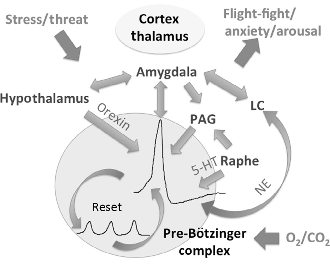
"Sighs, tears, grief, distress" expresses Johann Sebastian Bach in a musical example for the relationship between sighs and deep emotions. This review explores the neurobiological basis of the sigh and its relationship with psychology, physiology, and pathology. Sighs monitor changes in brain states, induce arousal, and reset breathing variability. These behavioral roles homeostatically regulate breathing stability under physiological and pathological conditions. Sighs evoked in hypoxia evoke arousal and thereby become critical for survival. Hypoarousal and failure to sigh have been associated with sudden infant death syndrome. Increased breathing irregularity may provoke excessive sighing and hyperarousal, a behavioral sequence that may play a role in panic disorders. Essential for generating sighs and breathing is the pre-Bötzinger complex. Modulatory and synaptic interactions within this local network and between networks located in the brainstem, cerebellum, cortex, hypothalamus, amygdala, and the periaqueductal gray may govern the relationships between physiology, psychology, and pathology. Unraveling these circuits will lead to a better understanding of how we balance emotions and how emotions become pathological.

Keywords: PAG; SIDS; anxiety; arousal; breathing; cardiorespiratory; panic; pre-Bötzinger complex; rhythm generation.


Figures

Figure 1
Figure 1
The neuronal, anatomical, and physiological characteristics of the sigh and the pre-Bötzinger complex in an intact animal, in a human, and in an in vitro preparation. The sigh recorded as integrated phrenic nerve activity from an intact animal (A) and as integrated population activity (D) within the pre-Bötzinger complex (B and C), isolated in a transverse slice from the ventrolateral medulla of a mouse (B). (A) The sigh is characterized by a large inspiratory burst of activity (A1) that is triggered from a normal eupneic breath and that is followed by an apnea (A2) alsoreferred to as “postsigh apnea.” (B) Rhythmically active transverse slice: the right side of the slice depicts an activity map of inspiratory activity. In this activity map, red represents the location of maximal integrated neuronal inspiratory activity generated during the sigh, which overlaps with the site of the pre-Bötzinger complex. (C) The pre-Bötzinger complexin a human. Note the close proximity to the Nucleus ambiguous (NA), which contains cardiac vagal neurons that are responsible for the generation of parasympathetic activity in the heart. The close proximity of NA and the pre-Bötzinger complex presumably plays a role in the cardiorespiratory coupling. (D) Integrated population activity recorded from the pre-Bötzinger complex. Note that the large-amplitude burst is followed by a period of apnea. (For interpretation of the references to color in this figure legend, the reader is referred to the online version of this chapter.) Modified from (B) Lieske et al. (2000), (C) Schwarzacher et al. (2010), and (D) Lieske et al. (2000).
Figure 2
Figure 2
Sighs occur spontaneously in humans. In addition, the neuronal activity critical for the generation of the sigh can be recorded as a “fictive sigh” when the underlying network is isolated in a brainstem slice preparation from a mouse. (A) Sighs are recorded as large-amplitude breathing movements using inductance plethysmography bands in a young infant. Note the regular occurrence of the large deflections that represent sighs. Inset marked by a red arrow: Each sigh consists of a large-amplitude inspiratory effort that is triggered by a eupneic breath and is followed by a short period of apnea. (B) Fictive sighs occur also spontaneously in the pre-Bötzinger complex isolated in slices obtained from neonatal mice. Note that the recordings in the human infant (A) and isolated slice (B) have the same time scale, illustrating the remarkable regularity of the sigh rhythmic activity, which occurs at a much slower time scale than the eupneic activity. (For interpretation of the references to color in this figure legend, the reader is referred to the online version of this chapter.)
Figure 3
Figure 3
Cardiorespiratory coupling of a sigh recorded in a healthy human subject. The sigh recorded with an inductance plethysmography brand from the abdomen (upper trace) is characterized by a heart rate increase followed by a heart rate decrease shown here in the simultaneously recorded electrocardiogram (lower trace). (For the color version of this figure, the reader is referred to the online version of this chapter.)
Figure 4
Figure 4
Eupneic activity recorded from an intact animal and as a neuronal signal representing fictive eupneic activity in a transverse slice preparation. (A and B) Simultaneous intracellular recording from an inspiratory neuron within the pre-Bötzinger complex (upper trace) and integrated extracellularly recorded phrenic nerve activity (lower trace). (B) Simultaneous intracellular recording from an inspiratory neuron within the pre-Bötzinger complex (upper trace) and integrated extracellular population activity recorded of the pre-Bötzinger complex (lower trace). Note the remarkable similarities in the intracellularly recorded discharge pattern of the inspiratory neuron both in vivo (A) and in vitro (B). (For the color version of this figure, the reader is referred to the online version of this chapter.)
Figure 5
Figure 5
Pacemaker activity recorded within the pre-Bötzinger complex. (A) Intracellular recording of a synaptically isolated pacemaker neuron in which bursting depends on the activation of the CAN current. (B) Intracellular recording of a synaptically isolated pacemaker neuron in which bursting is driven by the persistent sodium current. (C) The discharge pattern of respiratory neurons within the pre-Bötzinger complex follows a gradient. Some neurons are more bursting (right side) than others. Many neurons are weakly bursting and some are tonically active. This gradient illustrates that pacemaker neurons form not a discrete population. (For the color version of this figure, the reader is referred to the online version of this chapter.) Modified from (A and B) Pena et al. (2004) and (C) Carroll and Ramirez (2013).
Figure 6
Figure 6
Role of neuromodulators in determining the bursting characteristics of respiratory neurons. (A) Substance P turns a weakly burstingneuron into a strongly bursting pacemaker neuron. (B) Oxotremorine, an acetylcholine agonist, inhibits bursting in an INap-dependent pacemaker (see hyperpolarization) and induces the generation of a large-amplitude burst. As the modulatory effect weakens, small and large bursts overlap. (For the color version of this figure, the reader is referred to the online version of this chapter.)
Figure 7
Figure 7
The respiratory network in the pre-Bötzinger complex shows a gradient of discharge pattern as illustrated in the multiarray electrode recording (I). (I) The simultaneous multiarray recording from 11 neurons (see ordinate 1–11 in A) represents the cycle-by-cycle spiking behavior for each neuron within a respiratory cycle. Each block in A shows the spike time of a different neuron relative to the onset of inspiratory activity. In addition, the instantaneous spike rates are coded as a heat map from 0 to 55 Hz. C represents the average discharge for each neuron, and in D, the overlay of two pairs of averaged discharge activity shows that neurons have slightly different activation patterns. (II) The onset of discharge relative to the population activity is very variable as shown here for an intracellularly recorded pacemaker neuron. (For the color version of this figure, the reader is referred to the online version of this chapter.) Modified from Carroll et al. (2013).
Figure 8
Figure 8
Neuromodulators can differentially modulate eupneic and sigh activity, characterized as network activity in a functional brainstem slice preparation. The acetylcholine agonist oxotremorine specifically inhibits eupneic activity and activates sigh activity in the isolated pre-Bötzinger complex. (For the color version of this figure, the reader is referred to the online version of this chapter.) Modified from Tryba et al. (2008).
Figure 9
Figure 9
The sigh has important roles in the control of arousal and in resetting breathing variability. The schematic represents some of the network interactions involved in centrally linking the sigh with areas such as the hypothalamus, amygdala, the locus ceruleus (LC), periaqueductal grey (PAG), and the Raphe nucleus involved in the control of the flight-fight response. The cortex and thalamus interact with these areas in a complex manner; the details are not depicted in this illustration for reasons of simplicity, but the reader is referred to the text for more details. Many of the interactions are reciprocal and function via the release of neuromodulators such as orexin, serotonin (5-HT), and norepinephrine (NE) acting on different receptor subtypes. (For the color version of this figure, the reader is referred to the online version of this chapter.)

References

    1. Abelson JL, Nesse RM, Weg JG, Curtis GC. Respiratory psychophysiology and anxiety: cognitive intervention in the doxapram model of panic. Psychosom. Med. 1996;58:302–313. - PubMed
    1. Abelson JL, Weg JG, Nesse RM, Curtis GC. Persistent respiratory irregularity in patients with panic disorder. Biol. Psychiatry. 2001;49:588–595. - PubMed
    1. Abelson JL, Khan S, Lyubkin M, Giardino N. Respiratory irregularity and stress hormones in panic disorder: exploring potential linkages. Depress. Anxiety. 2008;25:885–887. - PubMed
    1. Abubakr A, Ifeayni I, Wambacq I. The efficacy of routine hyperventilation for seizure activation during prolonged video-electroencephalography monitoring. J. Clin. Neurosci. 2010;17:1503–1505. - PubMed
    1. Altmann LJ, Troche MS. High-level language production in Parkinson’s disease: a review. Parkinsons Dis. 2011;2011:238956. - PMC - PubMed

Publication types

LinkOut - more resources