

Analysis and validation of tissue biomarkers for renal cell carcinoma using automated high-throughput evaluation of protein expression
- PMID: 24746216
- PMCID: PMC4134351
- DOI: 10.1016/j.humpath.2014.01.008
Analysis and validation of tissue biomarkers for renal cell carcinoma using automated high-throughput evaluation of protein expression
Abstract
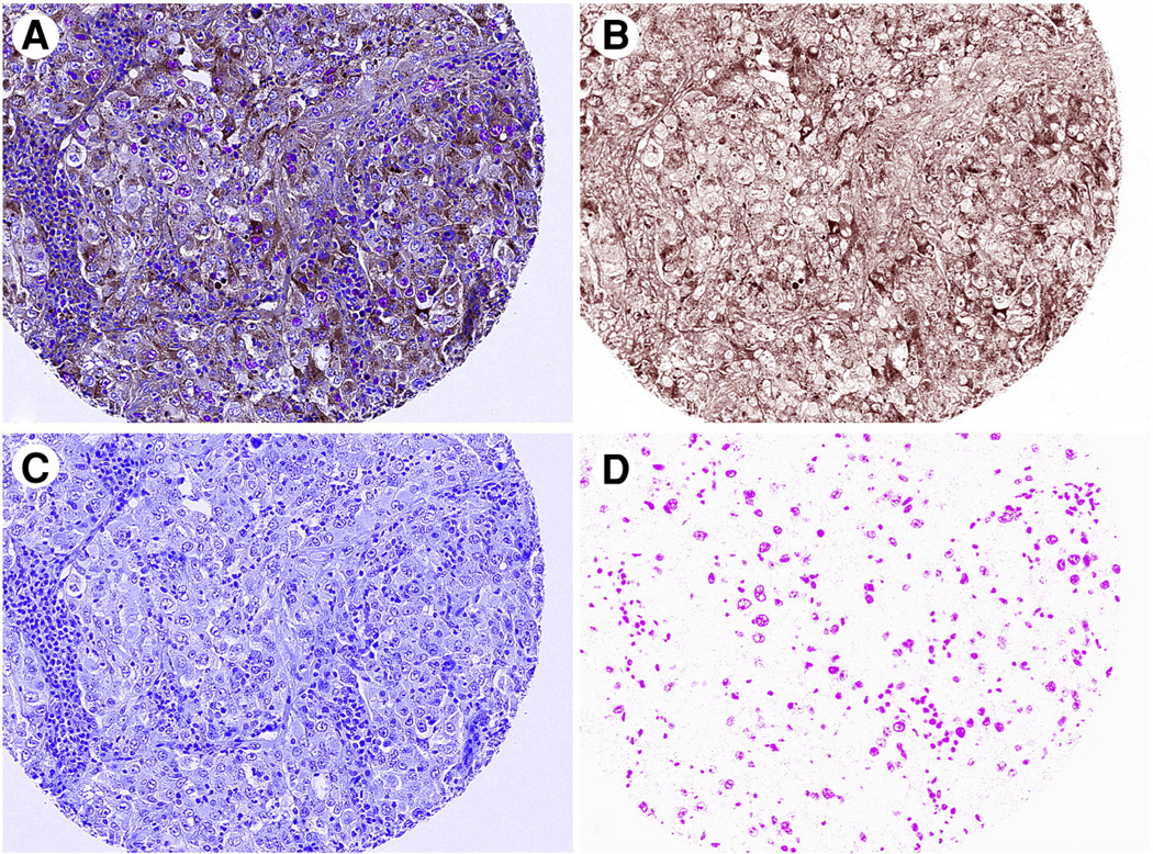
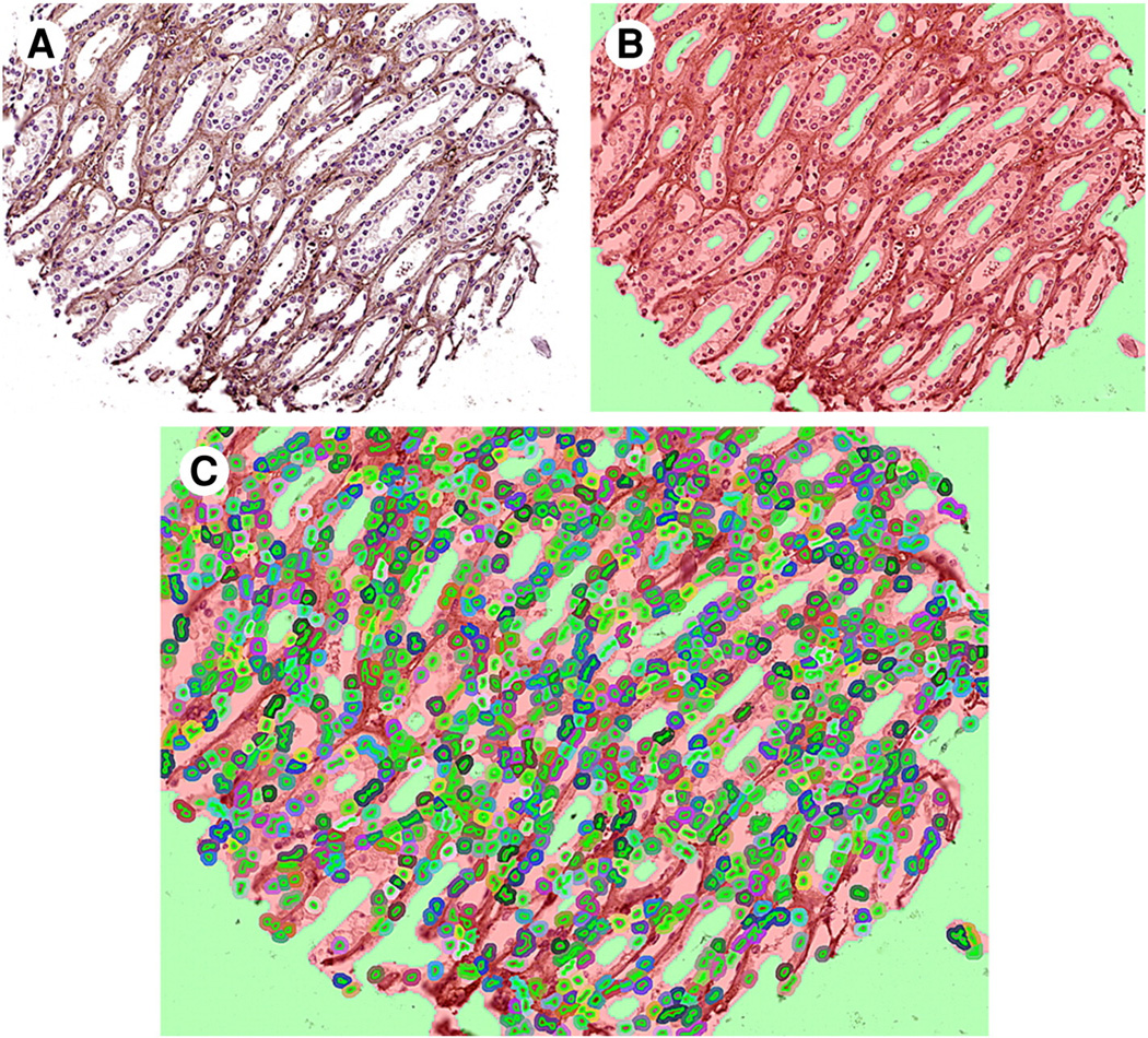
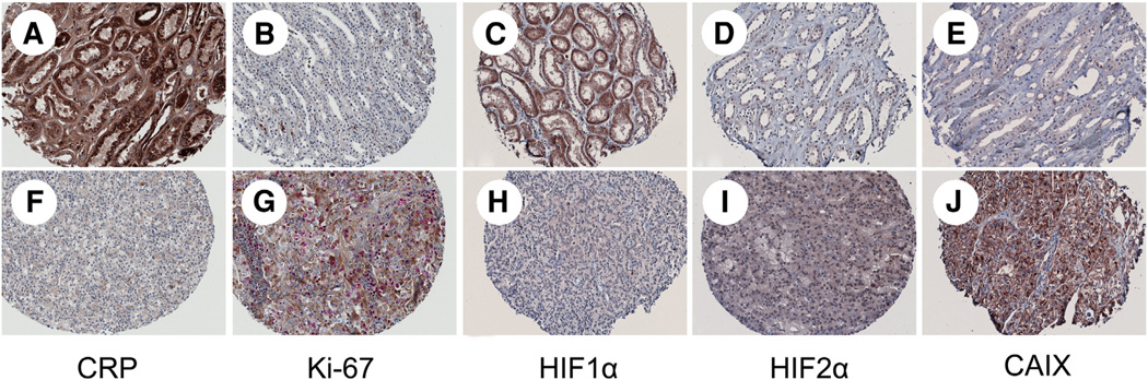
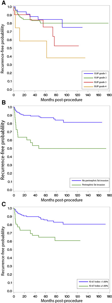
The objective of this study was to compare the predictive ability of potential tissue biomarkers to known prognostic factors that predict renal cell carcinoma (RCC) recurrence using an automated system of immunohistochemical analysis. After institutional review board approval, a tissue microarray was constructed using tissue from patients who had partial or radical nephrectomy for RCC. Patients with metastatic disease were excluded. Immunohistochemical staining of the tissue microarray for Ki-67, C-reactive protein, carbonic anhydrase 9, and hypoxia-inducible factors 1α and 2α was analyzed using automated image analysis. Univariable and multivariable analyses were performed to evaluate the association of putative biomarkers and known prognostic factors. Of 216 patients who met the entrance criteria, 34 (16%) patients developed metastatic recurrence within a median follow-up interval of 60.9 (interquartile range, 13.9-87.1) months. RCC morphotypes analyzed in this study include clear cell (n = 156), papillary (n = 38), chromophobe (n = 16), and collecting duct/unclassified (n = 6). Univariate analysis identified that only increased Ki-67 was predictive of RCC recurrence among the proteins evaluated, in addition to other known clinicopathological prognostic factors. After multivariate analysis, Ki-67 was identified as an independently predictive risk factor for RCC recurrence (hazard ratio [HR], 3.73 [confidence interval {CI}, 1.60-8.68]). Other independent predictors of RCC recurrence included tumor diameter (HR, 1.20 [CI, 1.02-1.41]) and perinephric fat invasion (HR, 4.49 [CI, 1.11-18.20]). We conclude that Ki-67 positivity is independently predictive of RCC recurrence after surgery in nonmetastatic patients. Automated analysis of tissue protein expression can facilitate a more objective and expedient investigation of tissue biomarkers for RCC.
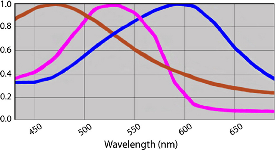
Keywords: Biomarkers; Immunohistochemistry; Multispectral imaging; Recurrence; Renal cell carcinoma.
Copyright © 2014 Elsevier Inc. All rights reserved.
Figures
References
-
- Siegel R, Naishadham D, Jemal A. Cancer statistics, 2013. CA Cancer J Clin. 2013;63:11–30. - PubMed
-
- Dudderidge TJ, Stoeber K, Loddo M, et al. Mcm2, Geminin, and Ki67 define proliferative state and are prognostic markers in renal cell carcinoma. Clin Cancer Res. 2005;11:2510–2517. - PubMed
-
- Sun M, Shariat SF, Cheng C, et al. Prognostic factors and predictive models in renal cell carcinoma: a contemporary review. Eur Urol. 2011;60:644–661. - PubMed
-
- Rimm DL. C-Path: a Watson-like visit to the pathology lab. Sci Transl Med. 2011;3:108fs8. - PubMed
-
- Huang WH, Kenneth H, Drew S. A colorful future of quantitative pathology: validation of Vectra technology using chromogenic multiplexed immunohistochemistry and prostate tissue microarrays. Hum Pathol. 2012;44:29–38. - PubMed
Publication types
MeSH terms
Substances
Grants and funding
LinkOut - more resources
Full Text Sources
Other Literature Sources
Research Materials
Miscellaneous
