Review
. 2014 May;25(5):225-34.
doi: 10.1016/j.tem.2014.03.010. Epub 2014 Apr 17.

Self-eating in the plaque: what macrophage autophagy reveals about atherosclerosis

Affiliations
Review

Self-eating in the plaque: what macrophage autophagy reveals about atherosclerosis

Ismail Sergin et al. Trends Endocrinol Metab. 2014 May.

Abstract

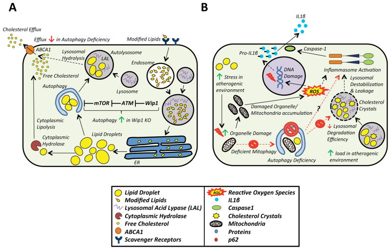
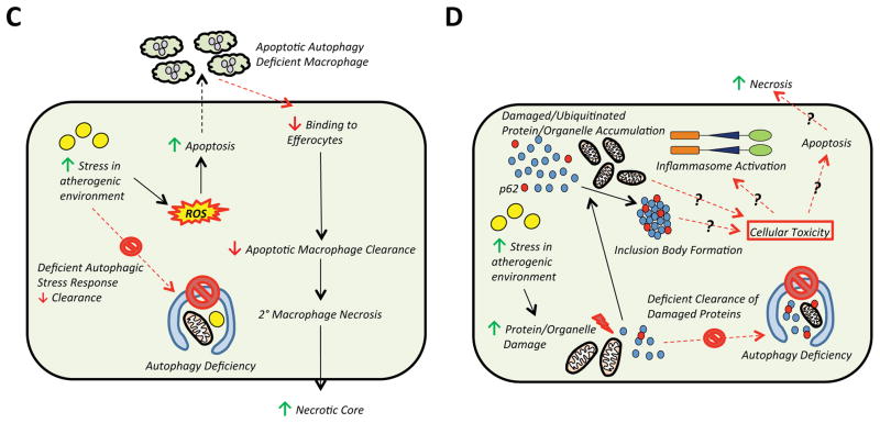
Autophagy (or 'self-eating') is the process by which cellular contents are recycled to support downstream metabolism. An explosion in research in the past decade has implicated its role in both health and disease and established the importance of the autophagic response during periods of stress and nutrient deprivation. Atherosclerosis is a state where chronic exposure to cellular stressors promotes disease progression, and alterations in autophagy are predicted to be consequential. Recent reports linking macrophage autophagy to lipid metabolism, blunted inflammatory signaling, and an overall suppression of proatherogenic processes support this notion. We review these data and provide a framework for understanding the role of macrophage autophagy in the pathogenesis of atherosclerosis, one of the most formidable diseases of our time.

Keywords: atherosclerosis; autophagy; inflammation; lipid metabolism; macrophage.


Figures

Box 1, Figure I
Box 1, Figure I. Overview of Autophagy
Autophagy begins with the formation of a double-membrane vesicle (autophagosome) which engulfs the adjacent cytoplasm including proteins and organelles. More than 30 proteins serve as the machinery to promote autophagosome nucleation/formation, vesicle elongation, and eventual docking/fusion with the lysosome. Certain autophagy proteins have been studied extensively and served as targets for mouse models of autophagy deficiency; these include Beclin-1 (involved in vesicle nucleation) and ATG5 and ATG7 (involved in vesicle elongation). Although autophagy is a bulk degradation process, chaperone proteins such as p62/SQSTM1 can selectively shuttle polyubiquitinated proteins and organelles for degradation (so-called selective autophagy). Autophagy is capable of being induced in response to various cellular stresses, the classic one being nutrient deprivation. Accordingly, mTOR, a major nutrient sensing kinase in cells, is a critical regulator of autophagy. Upon formation of the vesicle, autophagosomes fuse with lysosomes to generate autolysosomes, exposing the internal contents to lysosomal hydrolases for degradation into the basic metabolites needed for cellular function.
Figure 1
Figure 1. The Role of Monocytes/Macrophages in Atherosclerotic Progression
Atherosclerosis starts with the accumulation of lipid molecules/lipoproteins in the intima, which under normal circumstances is a small region below the endothelial layer (1). Activation of the endothelium leads to cytokine secretion, recruitment of circulating monocytes, and differentiation into macrophages (2). Macrophages uptake the accumulated lipids by receptor- mediated endocytosis. This includes both native and modified lipids such as oxidized LDL which are internalized by scavenger receptors (3). Lipid-exposed macrophages exacerbate the oxidative environment accelerating the lipid modification and uptake process. Macrophages also secrete inflammatory cytokines which lead to further monocyte entry to the region and local macrophage proliferation (4). Macrophage accumulation along with increased extracellular matrix production are major contributors to intimal expansion (4,5). When lipid overload exceeds the metabolic capacity of macrophages, intracellular lipid accumulation occurs leading to macrophage foam cell formation (6). Foam cell apoptosis and inefficient clearance further activate inflammation, intimal growth, and necrotic core formation, thereby contributing to increased lesion complexity and risk of rupture.
Figure 2
Figure 2. Role of Autophagy in Atherosclerotic Macrophages and the Consequences of Impaired Autophagy
(Left side) Macrophages located in the atherosclerotic plaque are subject to an environment of lipid excess. The autophagy-lysosomal system plays a central role in the response to such overload. The increased burden of cytoplasmic lipid droplets as well as damaged organelles and proteins are carried to the lysosomes by autophagy. The ensuing degradation not only prevents the accumulation of cytotoxic cargo, but also serves as an important mechanism for cholesterol efflux. (Right side) An impairment in autophagy results in several known consequences. (1) Cholesterol efflux decreases with a (2) concomitant accumulation of intracellular lipids. (3) Inflammasomes are hyperactivated leading to increased IL-1β secretion. (4) Cells are prone to apoptotic death while their clearance by efferocytosis is less efficient. (5) Inefficient degradation leads to the accumulation of damaged organelles and proteins. Taken together, these events exacerbate macrophage dysfunction and plaque progression.
Figure 3
Figure 3. Mechanisms That Link Macrophage Autophagy to Atherosclerosis
A) Although lipolysis by cytoplasmic hydrolyses were thought to be the only mechanism for cholesterol efflux, the role of autophagy in cholesterol efflux is now appreciated. Neutral lipid stores, a prominent feature of foam cells, are engulfed by autophagosomes via lipophagy, shuttled to lysosomes, hydrolyzed by lysosomal acid lipase, and the freed cholesterol is effluxed to the periphery via an ABCA1-dependent mechanism. In autophagy deficiency, the capacity of macrophages to efflux cholesterol efflux diminishes significantly. Pathways which regulate autophagy also have an impact on cholesterol efflux. Wip1 phosphatase, which activates mTOR in an ATM-dependent manner, is such an example. Wip1 KOs show decreased mTOR activation, increased lipophagy, and cholesterol efflux. B) Deficient autophagy also induces inflammasome activation and IL-1β secretion. The mechanistic link between autophagy deficiency and increased inflammasome activation is not clear. Possibilities include the accumulation of damaged mitochondria due to inefficient mitophagy with subsequent increases in ROS. ROS can induce inflammasome activation either directly or indirectly via its ability to cause DNA damage and secondary inflammatory signaling. Autophagy deficiency also reduces lysosomal degradation efficiency. The accumulation of intralysosomal lipids and cholesterol crystals is accelerated with autophagy deficiency, which may cause lysosomal membrane destabilization, lysosomal leakage, and inflammasome activation. C) Defects in autophagy lead to increased ROS and apoptotic cell death. Clearance of apoptotic autophagy-deficient macrophages by efferocytosis is also abrogated through unclear mechanisms. The accumulation of apoptotic and dying cells causes secondary necrosis which increases the necrotic core and lesion size and complexity. D) Although not specifically examined in atherosclerotic models, it is known that autophagy deficiency is also linked to the accumulation of damaged proteins/organelles and p62/SQSTM1-enriched inclusion body formation. Levels of p62/SQSTM1 are known to be markedly elevated in atherosclerotic plaques, suggesting that this aggregate formation is not only a marker of autophagy deficiency but might mediate cellular toxicity, inflammasome activation, and apoptosis as observed in other disease conditions where protein aggregation occurs.

References

    1. Go AS, et al. Executive summary: heart disease and stroke statistics--2013 update: a report from the American Heart Association. Circulation. 2013;127(1):143–52. - PubMed
    1. Williams KJ, Tabas I. The response-to-retention hypothesis of early atherogenesis. Arteriosclerosis, thrombosis, and vascular biology. 1995;15(5):551–61. - PMC - PubMed
    1. Moore KJ, Tabas I. Macrophages in the pathogenesis of atherosclerosis. Cell. 2011;145(3):341–55. - PMC - PubMed
    1. Ley K, Miller YI, Hedrick CC. Monocyte and macrophage dynamics during atherogenesis. Arteriosclerosis, thrombosis, and vascular biology. 2011;31(7):1506–16. - PMC - PubMed
    1. Moore KJ, Sheedy FJ, Fisher EA. Macrophages in atherosclerosis: a dynamic balance. Nature reviews Immunology. 2013;13(10):709–21. - PMC - PubMed

LinkOut - more resources