. 2014 Jul 15;391(2):125-32.
doi: 10.1016/j.ydbio.2014.04.003. Epub 2014 Apr 16.

Resident progenitors, not exogenous migratory cells, generate the majority of visceral mesothelium in organogenesis

Affiliations

Resident progenitors, not exogenous migratory cells, generate the majority of visceral mesothelium in organogenesis

Nichelle I Winters et al. Dev Biol. .

Abstract

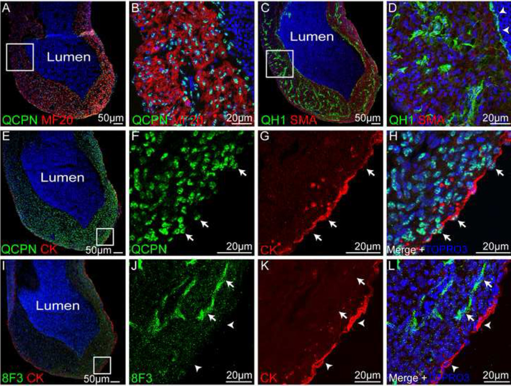
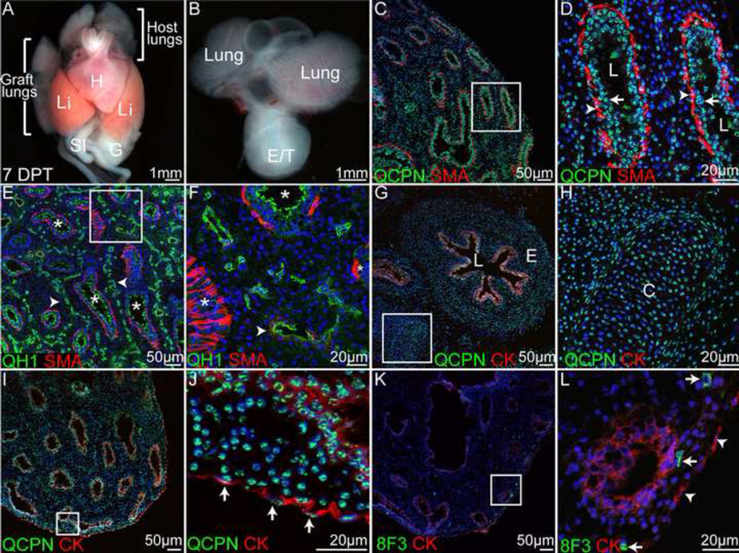
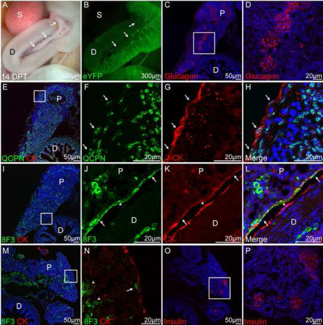
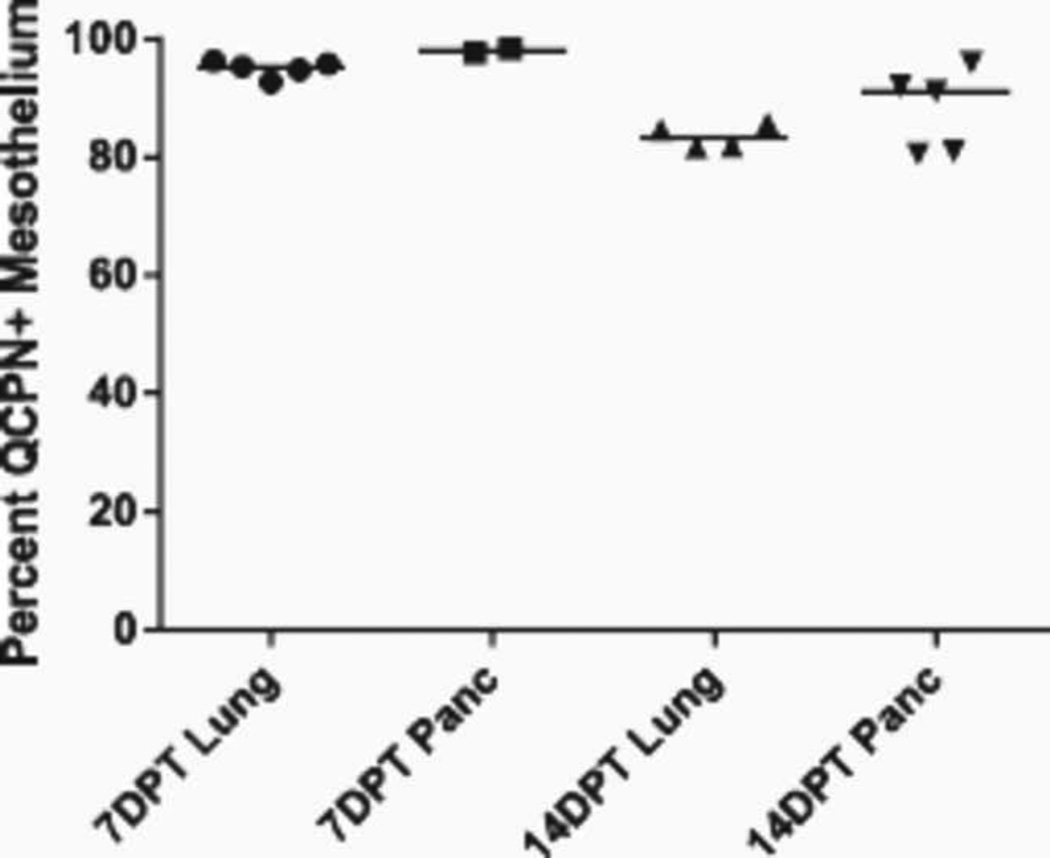
Historically, analyses of mesothelial differentiation have focused on the heart where a highly migratory population of progenitors originating from a localized "extrinsic" source moves to and over the developing organ. This model long stood alone as the paradigm for generation of this cell type. Here, using chick/quail chimeric grafting and subsequent identification of mesothelial cell populations, we demonstrate that a different mechanism for the generation of mesothelia exists in vertebrate organogenesis. In this newly discovered model, mesothelial progenitors are intrinsic to organs of the developing digestive and respiratory systems. Additionally, we demonstrate that the early heart stands alone in its ability to recruit an entirely exogenous mesothelial cell layer during development. Thus, the newly identified "organ intrinsic" model of mesotheliogenesis appears to predominate while the long-studied cardiac model of mesothelial development may be the outlier.

Keywords: Mesothelia; Organogenesis; coelomate.


Figures

Figure 1
Figure 1. Quail heart tube isolated without proepicardium 7DPT
Representative 7DPT graft-derived heart. A) Graft developed within the coelomic cavity of the host in close proximity to the body wall (BW), liver (Li) and kidney (K) and was tube like in shape (arrow). B) Whole mount eYFP fluorescence. A blood vessel (arrowhead) extended from the graft (arrows) to the host. C) Quail cells (QCPN-positive) within the graft are myosin (MF20)-positive suggestive of cardiomyocyte differentiation. D) Neighboring QCPN-negative cells are also negative for MF20 (arrowheads). E) Low power image demonstrating myosin staining within the graft-derived heart. F) Higher power of boxed area in E). G) Higher power of boxed area in F) demonstrates striations consistent with sarcomere formation (arrows). H) Endocardial/endothelial cells marked by the antibody QH1 were found within the graft (arrows). I) Graft-derived heart was covered by a cytokeratin-positive surface mesothelial layer. J–L) Higher power of boxed area in I). The surface mesothelium was QCPN-negative (arrowheads) and had an underlying mesenchymal layer of cells (asterisks). M) The graft-heart was surrounded on all sides by chick cells (8F3 positive). N–P) Higher magnification of boxed area in M). 8F3 co-localized with cytokeratin demonstrating the host origin of the graft mesothelium. BW, body wall; H, heart; K, kidney; Li, liver; W, wing.
Figure 2
Figure 2. Quail heart tube isolated with attached proepicardium/epicardium 7DPT
Heart tubes were isolated from HH19–20 quail embryos after the PE had attached and the epicardium had partially formed. A) QCPN-positive myocytes encircled a blood filled lumen. B) Higher magnification of boxed area in A. C) Quail endothelial/endocardial cells (QH1-positive) were distributed throughout the graft-derived heart. D) Higher magnification of boxed area in C. Endocardial cells lined the lumen (arrowheads). E) Low power view demonstrating cytokeratinpositive mesothelium lining the surface of the graft. F–H) Higher magnification of boxed area in E). The surface mesothelial cells were QCPN-positive (arrows). I) Low power view demonstrating scattered chick (8F3-positive) cells within the graft-derived heart tube. J–L) Higher magnification of boxed area in I). Chick cells were found within the graft (arrows) but not within the mesothelium (arrowhead).
Figure 3
Figure 3. Graft-derived lungs 7DPT
A) Transplanted lung buds formed paired lungs within the coelomic cavity of the host embryo (H, heart, Li, liver, SI, small intestine, G, gizzard). B) A central esophagus/trachea (E/T) also formed from the grafted tissue. C) The graft-derived lungs were formed primarily of quail tissue and included a linear organization of airways. D) Higher magnification of boxed area in C) (L, lumen). The mucosal linings of airways were derived from quail (arrows) as was the encircling smooth muscle (arrowheads). E) Quail endothelial cells marked by QH1 surrounded the airways (arrowheads). The mucosal epithelium was also QH1-positive (asterisks) as previously reported (26). F) Higher magnification of boxed area in E). The major vessels exhibited limited vascular smooth muscle differentiation (arrowhead) and were localized centrally between airways (asterisks). G) Section through the lumen (L) of the graft-derived esophagus (E). H) Higher magnification of boxed area in G. Near the esophageal lumen, cartilage (C) differentiation was frequently observed. I) Low magnification demonstrating cytokeratin-positive surface epithelium. J) Surface mesothelium was QCPN-positive. K) Chick cells were scattered throughout the graft in low numbers. L) Chick cells (arrows) did not contribute to the mesothelium (arrowheads). E/T, esophagus/trachea; G, gizzard; H, heart; Li, liver; SI, small intestine.
Figure 4
Figure 4. Graft-derived pancreas 14 DPT
A) The grafted tissue formed a duodenum (D) and pancreas (arrows). B) Whole mount eYFP fluorescence. C) Within the graft-derived pancreas there were islands of glucagon positive cells (red). D) Higher magnification of boxed area in C). E) Both the pancreas and duodenum were formed in large part by QCPN-positive cells. F–H) Higher magnification of boxed area in E). The surface cells were QCPN- and cytokeratinpositive (arrows) demonstrating their quail origin. I) Chick cells were also distributed throughout the graft. J–L) Higher magnification of boxed area in I). Rare patches of surface mesothelial cells were 8F3-positive (arrows). The mesothelial cells of the nearby duodenum were 8F3-negative (arrowhead). M) Low power view of another graft-derived pancreas and duodenum. N) In this graft, 8F3 cells were not found within the mesothelium (arrow) and most were within clusters deep within the pancreas (arrowheads). O–P) This graft also exhibited islet formation with insulin production (red). D, duodenum; P, pancreas; S, spleen. See also Figure S4.
Figure 5
Figure 5. Quantification of QCPN-positive mesothelium in graft-derived organs
Each symbol represents a single experimental embryo. Horizontal line denotes mean value for each sample group. At 7DPT, lung and pancreatic graft mesothelia were between 93–98% QCPN-positive. At 14DPT, lung graft mesothelia were on average 84% QCPN-positive. Pancreatic graft mesothelia were on average 88% QCPN-positive at 14DPT.

References

    1. Que J, et al. Mesothelium contributes to vascular smooth muscle and mesenchyme during lung development. Proc Natl Acad Sci U S A. 2008;105(43):16626–16630. - PMC - PubMed
    1. Asahina K, Zhou B, Pu WT, Tsukamoto H. Septum transversum derived mesothelium gives rise to hepatic stellate cells and perivascular mesenchymal cells in developing mouse liver. Hepatology. 2011;53(3):983–995. - PMC - PubMed
    1. Wilm B, Ipenberg A, Hastie ND, Burch JB, Bader DM. The serosal mesothelium is a major source of smooth muscle cells of the gut vasculature. Development. 2005;132(23):5317–5328. - PubMed
    1. Dettman RW, Denetclaw W, Jr, Ordahl CP, Bristow J. Common epicardial origin of coronary vascular smooth muscle, perivascular fibroblasts, and intermyocardial fibroblasts in the avian heart. Dev Biol. 1998;193(2):169–181. - PubMed
    1. Li P, et al. IGF signaling directs ventricular cardiomyocyte proliferation during embryonic heart development. Development. 2011;138(9):1795–1805. - PMC - PubMed

Publication types

LinkOut - more resources