. 2014 Aug:73:51-9.
doi: 10.1016/j.freeradbiomed.2014.04.014. Epub 2014 Apr 16.

Inducible nitric oxide synthase is key to peroxynitrite-mediated, LPS-induced protein radical formation in murine microglial BV2 cells

Affiliations

Inducible nitric oxide synthase is key to peroxynitrite-mediated, LPS-induced protein radical formation in murine microglial BV2 cells

Ashutosh Kumar et al. Free Radic Biol Med. 2014 Aug.

Abstract

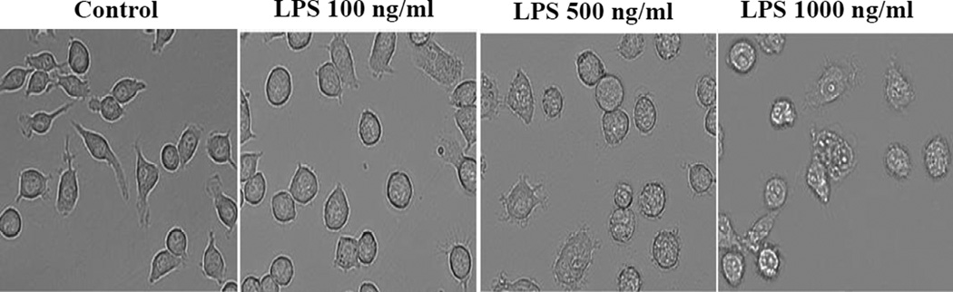
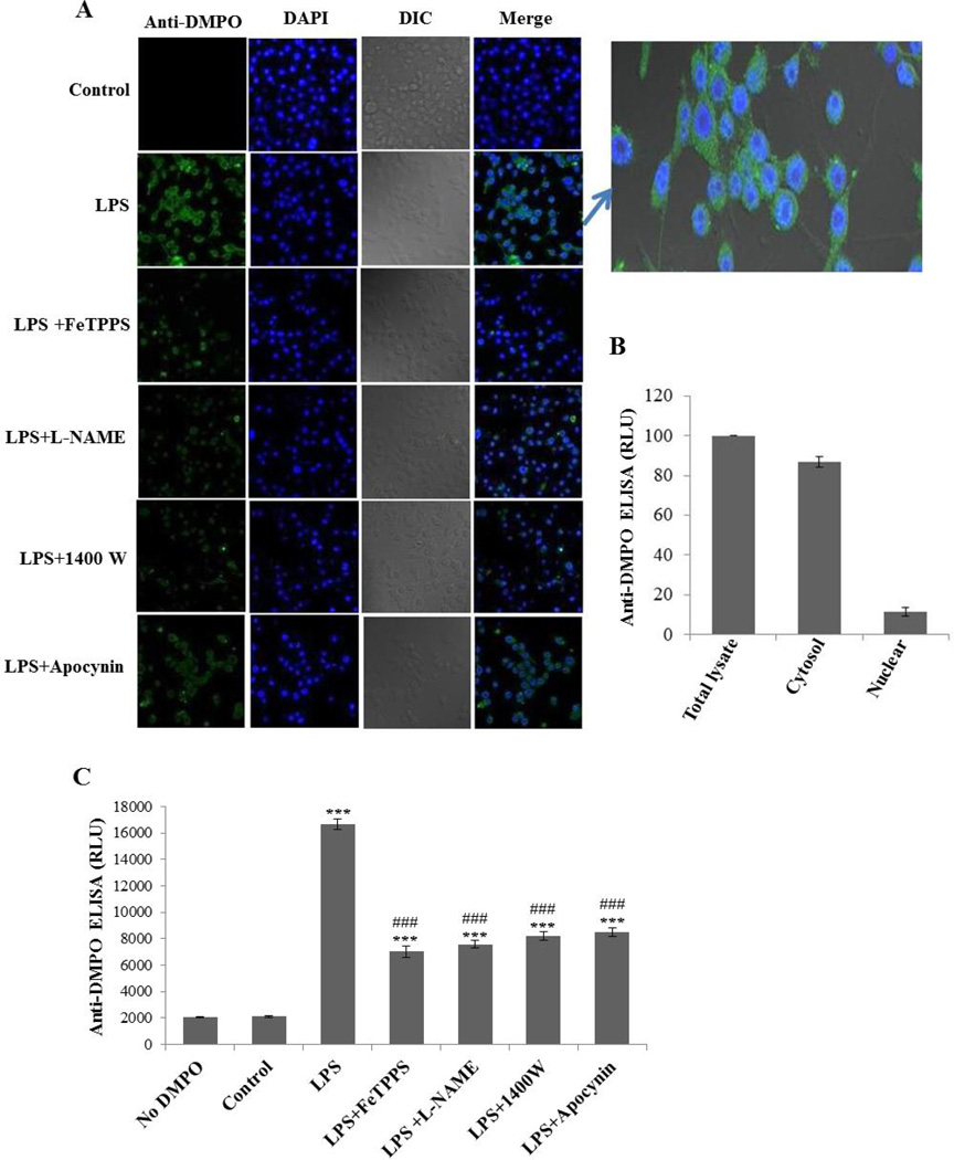
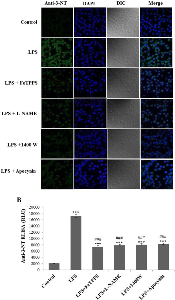
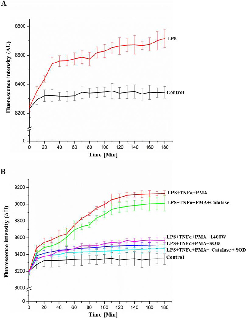
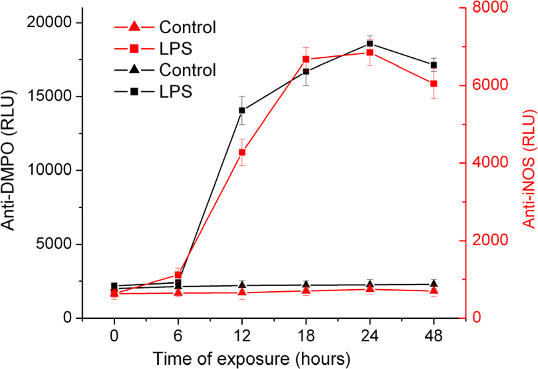
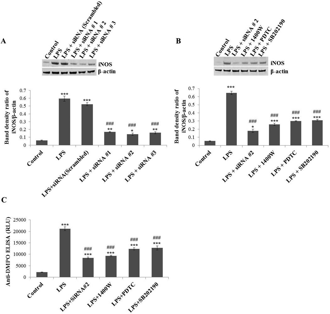
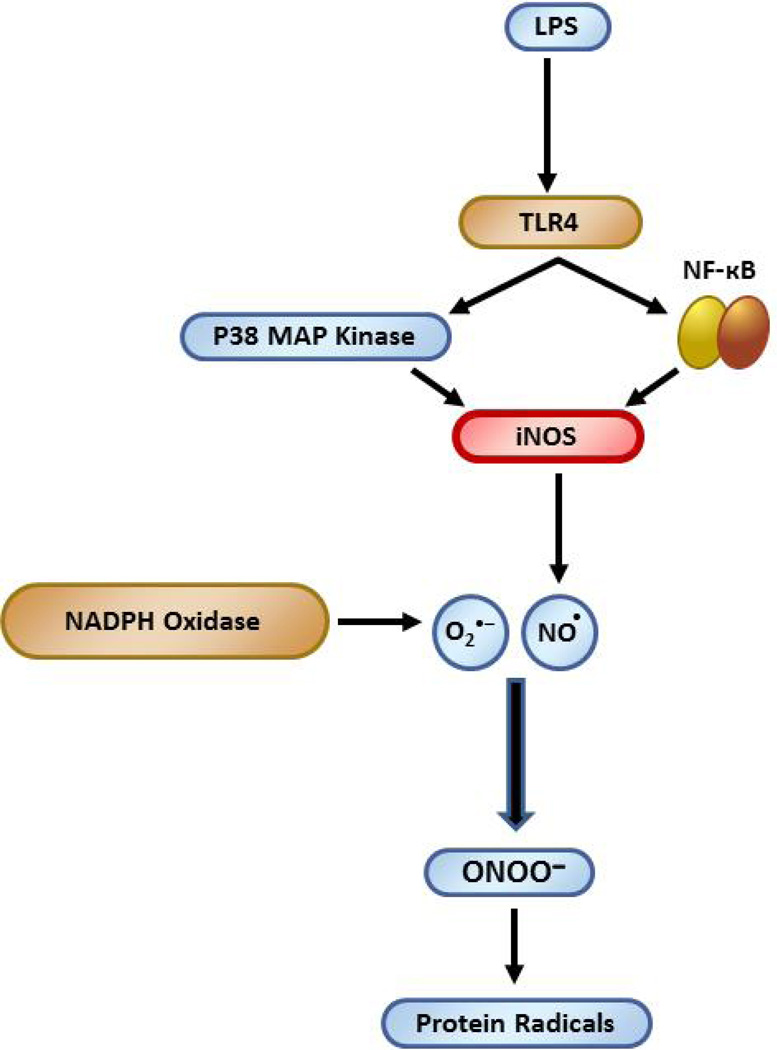
Microglia are the resident immune cells in the brain. Microglial activation is characteristic of several inflammatory and neurodegenerative diseases including Alzheimer's disease, multiple sclerosis, and Parkinson's disease. Though lipopolysaccharide (LPS)-induced microglial activation in models of Parkinson's disease is well documented, the free radical-mediated protein radical formation and its underlying mechanism during LPS-induced microglial activation are not known. Here we have used immuno-spin trapping and RNA interference to investigate the role of inducible nitric oxide synthase (iNOS) in peroxynitrite-mediated protein radical formation in murine microglial BV2 cells treated with LPS. Treatment of BV2 cells with LPS resulted in morphological changes, induction of iNOS, and increased protein radical formation. Pretreatments with FeTPPS (a peroxynitrite decomposition catalyst), L-NAME (total NOS inhibitor), 1400W (iNOS inhibitor), and apocynin significantly attenuated LPS-induced protein radical formation and tyrosine nitration. Results obtained with coumarin-7-boronic acid, a highly specific probe for peroxynitrite detection, correlated with LPS-induced tyrosine nitration, which demonstrated involvement of peroxynitrite in protein radical formation. A similar degree of protection conferred by 1400W and L-NAME led us to conclude that only iNOS, and no other forms of NOS, is involved in LPS-induced peroxynitrite formation. Subsequently, siRNA for iNOS, the iNOS-specific inhibitor 1400W, the NF-κB inhibitor PDTC, and the p38 MAPK inhibitor SB202190 was used to inhibit iNOS directly or indirectly. Inhibition of iNOS precisely correlated with decreased protein radical formation in LPS-treated BV2 cells. The time course of protein radical formation also matched the time course of iNOS expression. Taken together, these results prove the role of iNOS in peroxynitrite-mediated protein radical formation in LPS-treated microglial BV2 cells.

Keywords: Free radicals; Inducible nitric oxide synthase; Lipopolysaccharide; Microglia; Nitrone adducts; Parkinson disease; Peroxynitrite; Protein radical.


Conflict of interest statement

Conflict of interest: The authors declare no conflict of interest.

Figures

Figure 1
Figure 1
LPS triggers microglial activation and changes in morphology. Phase contrast images showing morphology of BV2 cells treated with different concentrations of LPS for 24 hours.
Figure 2
Figure 2
LPS induces protein radical formation in BV2 cells. (A) Confocal images showing the anti-DMPO staining of BV2 cells treated with 500 ng/ml LPS and 50 mM DMPO for 24 hours in the presence and absence of pretreatment with FeTPPS (10 µM), L-NAME (100 µM), 1400W (10 µM), and apocynin (1 mM). (B) Anti-DMPO ELISA from the nuclear and cytosolic fractions of LPS-treated BV2 cells. (C) Anti-DMPO ELISA of BV2 cells treated with 500 ng/ml LPS and 50 mM DMPO for 24 hours in the presence and absence of pretreatment with FeTPPS (10 µM), L-NAME (100 µM), 1400W (10 µM), and apocynin (1 mM). Data show mean values ± SEM or representative images from three independent experiments (n=6). (*** P<0.001, with respect to control, and ### P<0.001, with respect to the LPS-treated group.)
Figure 3
Figure 3
LPS induces tyrosine nitration in BV2 cells. (A) Confocal images showing anti-3-nitro-tyrosine staining of BV2 cells treated with 500 ng/ml LPS and 50 mM DMPO for 24 hours in the presence and absence of pretreatment with FeTPPS (10 µM), L-NAME (100 µM), 1400W (10 µM), and apocynin (1 mM). (B) Anti-nitrotyrosine ELISA of BV2 cells treated with 500 ng/ml LPS and 50 mM DMPO for 24 hours in the presence and absence of pretreatment with FeTPPS (10 µM), L-NAME (100 µM), 1400W (10 µM), and apocynin (1 mM). Data show mean values ± SEM or representative images from three independent experiments (n=6). (*** P<0.001, with respect to control, and ### P<0.001, with respect to the LPS-treated group.)
Figure 4
Figure 4
Measurement of peroxynitrite formation by monitoring the oxidation of coumarin-7-boronic acid. (A) Cells were incubated with LPS (500 ng/ml) for 21 hours and then CBA was added (20 µM) to the medium. (B) Cells were incubated with LPS (500 ng/ml) and TNFα (50 ng/ml) in the presence and absence of the iNOS inhibitor 1400W (10 µM) for 21 hours, and then co-treated with CBA and PMA (200 ng/ml) in the same medium. Additionally, in some samples, the O2 •− scavenger SOD (500 U/ml) and/or the H2O2 scavenger catalase (1000 U/ml) were added along with CBA and PMA. Continuous fluorescence intensity measurements were initiated immediately after CBA was added.
Figure 5
Figure 5
Correlation between iNOS expression and protein radical formation. Anti-DMPO and anti-iNOS ELISA of BV2 cells exposed to 500 ng/ml LPS and 50 mM DMPO (0–48 hours). Data show mean values ± SEM from three independent experiments (n=6).
Figure 6
Figure 6
Effect of iNOS inhibition/silencing on LPS-induced protein oxidation in BV2 cells. (A) BV2 cells were transfected with siRNA1, 2, 3 of iNOS or control (scrambled) siRNA. Forty-eight hours later, cells were incubated with LPS (500 ng/ml) for 24 hours and then harvested for Western blot. The upper panel shows a Western blot of iNOS and the lower panel shows the band density ratio of iNOS and β-actin. (B) BV2 cells transfected with most effective siRNA#2 for iNOS or cells exposed to direct (1400W) or indirect (PDTC, SB202190) inhibitors of iNOS were analyzed by Western blot (upper panel). The lower panel shows the band density ratio of iNOS and β-actin. (C) Anti-DMPO ELISA of BV2 cells treated with 500 ng/ml LPS and 50 mM DMPO for 24 hours in the presence and absence of iNOS silencing/inhibition. Data show mean values ± SEM or representative images from three independent experiments (n=6). (*P<0.05, ** P<0.01, *** P<0.001, with respect to control, and ### P<0.001, with respect to the LPS-treated group.
Scheme
Scheme
iNOS is crucial to LPS-induced protein radical formation in microglia Role of iNOS in LPS-induced protein radical formation in BV2 cells.

References

    1. Bezard E, Przedborski S. A tale on animal models of Parkinson's disease. Mov. Disord. 2011;26:993–1002. - PubMed
    1. Li M, Dai FR, Du XP, Yang QD, Chen Y. Neuroprotection by silencing iNOS expression in a 6-OHDA model of Parkinson's disease. J. Mol. Neurosci. 2012;48:225–233. - PubMed
    1. Purisai MG, McCormack AL, Cumine S, Li J, Isla MZ, Di Monte DA. Microglial activation as a priming event leading to paraquat-induced dopaminergic cell degeneration. Neurobiol. Dis. 2007;25:392–400. - PMC - PubMed
    1. Kreutzberg GW. Microglia: a sensor for pathological events in the CNS. Trends Neurosci. 1996;19:312–318. - PubMed
    1. Qin L, Liu Y, Hong JS, Crews FT. NADPH oxidase and aging drive microglial activation, oxidative stress, and dopaminergic neurodegeneration following systemic LPS administration. Glia. 2013;61:855–868. - PMC - PubMed

Publication types

MeSH terms

Substances