. 2014 May 8;54(3):407-417.
doi: 10.1016/j.molcel.2014.03.023. Epub 2014 Apr 17.

Fragile X mental retardation protein regulates translation by binding directly to the ribosome

Affiliations

Fragile X mental retardation protein regulates translation by binding directly to the ribosome

Eileen Chen et al. Mol Cell. .

Abstract

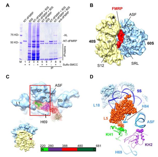
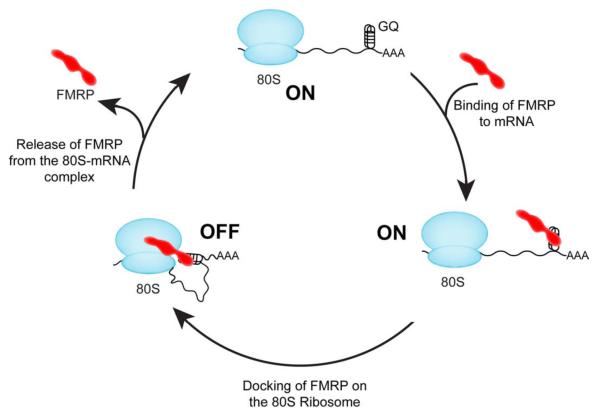
Fragile X syndrome (FXS) is the most common form of inherited mental retardation, and it is caused by loss of function of the fragile X mental retardation protein (FMRP). FMRP is an RNA-binding protein that is involved in the translational regulation of several neuronal mRNAs. However, the precise mechanism of translational inhibition by FMRP is unknown. Here, we show that FMRP inhibits translation by binding directly to the L5 protein on the 80S ribosome. Furthermore, cryoelectron microscopic reconstruction of the 80S ribosome⋅FMRP complex shows that FMRP binds within the intersubunit space of the ribosome such that it would preclude the binding of tRNA and translation elongation factors on the ribosome. These findings suggest that FMRP inhibits translation by blocking the essential components of the translational machinery from binding to the ribosome.


Figures

Figure 1
Figure 1. Inhibition of translation by FMRP
(A) Domain organization of dFMRP (1-681 amino acids) and NT-dFMRP (220-681 amino acids). NLS, nuclear localization signal; KH, K-homology domain; NES, nuclear export signal; RGG, motif rich in arginine and glycine. (B) Time course of luciferase mRNA translation. Red trace, control translation without NT-dFMRP; light blue trace, translation with 1.2 μM bovine serum albumin (BSA); cyan trace, translation with 1.2 μM RNA-binding MS2 coat protein; blue trace, translation with 0.6 μM NT-dFMRP. The addition of NT-dFMRP to the IVTS inhibited luciferase mRNA translation. In contrast, the addition of BSA or MS2 coat protein to the IVTS did not inhibit the synthesis of luciferase. (C) Inhibition of translation by NT-dFMRP mutants. Time course of luciferase mRNA translation in the presence of the indicated mutant NT-dFMRP proteins. (D) Inhibition of translation by FMRP is independent of WGGA and ACUK sequences. Red trace, control mRNA; green trace, mRNA without WGGA sequence; blue trace, mRNA without ACUK sequence; orange trace, mRNA without both WGGA and ACUK sequences. In all cases, the data were normalized with respect to the control translation without NT-dFMRP. The bar graphs next to each time course show the mean ± SD from three independent experiments. (E) Inhibition of translation by FMRP in cells. Translation of control luciferase mRNA (red bars) and luciferase mRNA without WGGA and ACUK sequences (orange bars) are inhibited to a similar extent by full-length dFMRP and NT-dFMRP, as indicated. Data were normalized with respect to control cells, which were co-transfected with an empty plasmid and the appropriate luciferase plasmid. The transfection experiments were done in duplicates and the mean ± SD from three independent transfection experiments are shown. (F) Inhibition of translation by FMRP is independent of G-quadruplex and pseudoknot forming sequences in the mRNA. Red trace, control mRNA; blue trace, mRNA with ΔKC2 pseudoknot forming sequence; green trace, mRNA with SC1 G-quadruplex sequence. The bar graph shows the mean ± SD from three independent experiments. See also Figures S1 to S4.
Figure 2
Figure 2. FMRP binds directly to the ribosome
(A) SDS-PAGE gel showing that NT-dFMRP elutes with the ribosome, whereas BSA remains in the gel filtration column. Lanes: M, molecular weight ladder; 1, ribosome only; 2, NT-dFMRP only; 3, ribosome + NT-dFMRP; 4, input NT-dFMRP directly loaded on the gel; 5, BSA only; 6, ribosome + BSA; 7, input BSA directly loaded on the gel. Positions of NT-dFMRP (51 KD, arrow) and BSA (67 KD) are indicated. Vertical black bar indicates ribosomal proteins. (B) KH1 domain of FMRP is important for binding to the ribosome. Lanes: M, molecular weight ladder; 1, ribosome only; 2, NT-dFMRP only; 3, ribosome + NT-dFMRP; 4, KH2 (I307N) mutant only; 5, KH2 ((I307N) mutant + ribosome; 6, KH1 (I244N) mutant only; 7, KH1 (I244N) mutant + ribosome. (C) Bar graph of ribosome binding data showing mean ± SD from three independent experiments.
Figure 3
Figure 3. FRET-based assay for determining the binding affinity of FMRP for the ribosome
(A) Schematic representation of the FRET assay. Ribosome (cyan) is labeled with Cy3 (yellow stars). NT-dFMRP (purple) is labeled with Cy5 (red star). The binding of NT-dFMRP to the ribosome results in FRET (emission at 665 nm). (B) Emission spectrum showing the changes in fluorescence intensity because of FMRP binding to the ribosome. Blue trace, 80S ribosome only control reaction; green trace, NT-dFMRP only control reaction; red trace, 80S ribosome + NT-dFMRP complex. Similar concentration of 80S ribosome and NT-dFMRP were used in the different reactions. (C) Binding curve for wild type NT-dFMRP. (D) Binding curve for KH1 (I244N) mutant. (E) Binding curve for KH2 (I307N) mutant. (F) Binding curve for the ΔRGG mutant. The y-axis shows the %FRET efficiency and the error bars show mean ± SD from three independent experiments.
Figure 4
Figure 4. FMRP binds near ribosomal protein L5
(A) SDS-PAGE gel showing that NT-dFMRP crosslinks to the ribosome. Lanes: M, molecular weight ladder; 1, NT-dFMRP only; 2, ribosome only; 3, ribosome + NT-dFMRP; 4, ribosome + NT-dFMRP. SMCC was added to samples in lanes 1, 2, and 4. Lanes 5 to 8 correspond to samples in lanes 1 to 4 after immunoprecipitation (IP) with anti-dFMRP antibody and purification. Lane 9 shows the heavy and light chains of the anti-dFMRP antibody. Black bar, ribosomal proteins; XL, position of the crosslinked proteins. (B) Isolated density corresponding to NT-dFMRP (red) superimposed onto the 11.2 Å resolution segmented cryo-EM map of the control Drosophila 80S ribosome (40S subunit, yellow; 60S subunit, blue) (see Figure S6 for a stereo representation of the FMRP binding region). (C) A portion of the 60S subunit map is shown from its interface side to reveal the overall binding position of NT-dFMRP. The fitted I-TASSER model of the NT-dFMRP is displayed onto the 60S subunit with fitted X-ray coordinates of the yeast 60S ribosome. Four structural domains of the NT-dFMRP are identified according to the color in the bar-diagram shown at the bottom. Thumbnail at lower left depicts the overall orientation of the ribosome in panels C and D. (D) The boxed area in panel C is enlarged, with cryo-EM densities removed and a cutting plane applied on the far side, to reveal putative interactions of KH1 and KH2 domains of NT-dFMRP with the 60S subunit components. Landmarks of the 40S subunit in panel b: hd, head; sh, shoulder, and S12, protein S12 region; and an asterisk (*) points to a Drosophila specific expansion sequence emerging from helix 39 of the 18S rRNA. Landmarks of the 60S subunit: L1, L1 protein protuberance; CP, central protuberance; Sb, L7/L12 stalk base; 5S, 5S rRNA; H69 and H84, 28S rRNA helices 69 and 84, respectively; ASF, A-site finger, or 28S rRNA helix 38; SRL, α-sarcin-ricin stem-loop; and 60S ribosomal proteins L5 and L18 are shown as space-filled models. See also Figures S5 to S7.
Figure 5
Figure 5. Schematic representation of translational inhibition by FMRP
FMRP (red) uses its RGG domain to bind to mRNAs having G-quadruplex (GQ) forming sequence and then docks on the 80S ribosome (blue) using the KH1 and KH2 domains. Binding of FMRP to the mRNA and the ribosome synergistically inhibit translation (OFF state). Activation of the neuron changes the post-translational modification status of FMRP, which releases FMRP from the 80S•mRNA complex and activates translation (ON state).

Comment in

References

    1. Adams-Cioaba MA, Guo Y, Bian C, Amaya MF, Lam R, Wasney GA, Vedadi M, Xu C, Min J. Structural studies of the tandem Tudor domains of fragile X mental retardation related proteins FXR1 and FXR2. PLoS One. 2010;5:e13559. - PMC - PubMed
    1. Agrawal RK, Spahn CM, Penczek P, Grassucci RA, Nierhaus KH, Frank J. Visualization of tRNA movements on the Escherichia coli 70S ribosome during the elongation cycle. J Cell Biol. 2000;150:447–460. - PMC - PubMed
    1. Agulhon C, Blanchet P, Kobetz A, Marchant D, Faucon N, Sarda P, Moraine C, Sittler A, Biancalana V, Malafosse A, et al. Expression of FMR1, FXR1, and FXR2 genes in human prenatal tissues. Journal of neuropathology and experimental neurology. 1999;58:867–880. - PubMed
    1. Ascano M, Jr., Mukherjee N, Bandaru P, Miller JB, Nusbaum JD, Corcoran DL, Langlois C, Munschauer M, Dewell S, Hafner M, et al. FMRP targets distinct mRNA sequence elements to regulate protein expression. Nature. 2012;492:382–386. - PMC - PubMed
    1. Ashley CT, Jr., Wilkinson KD, Reines D, Warren ST. FMR1 protein: conserved RNP family domains and selective RNA binding. Science. 1993;262:563–566. - PubMed

Publication types

MeSH terms