. 2014 May 8;7(3):796-806.
doi: 10.1016/j.celrep.2014.03.040. Epub 2014 Apr 17.

proBDNF negatively regulates neuronal remodeling, synaptic transmission, and synaptic plasticity in hippocampus

Affiliations

proBDNF negatively regulates neuronal remodeling, synaptic transmission, and synaptic plasticity in hippocampus

Jianmin Yang et al. Cell Rep. .

Abstract

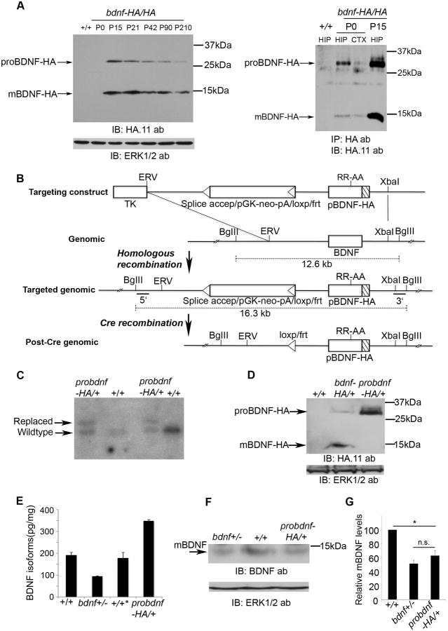
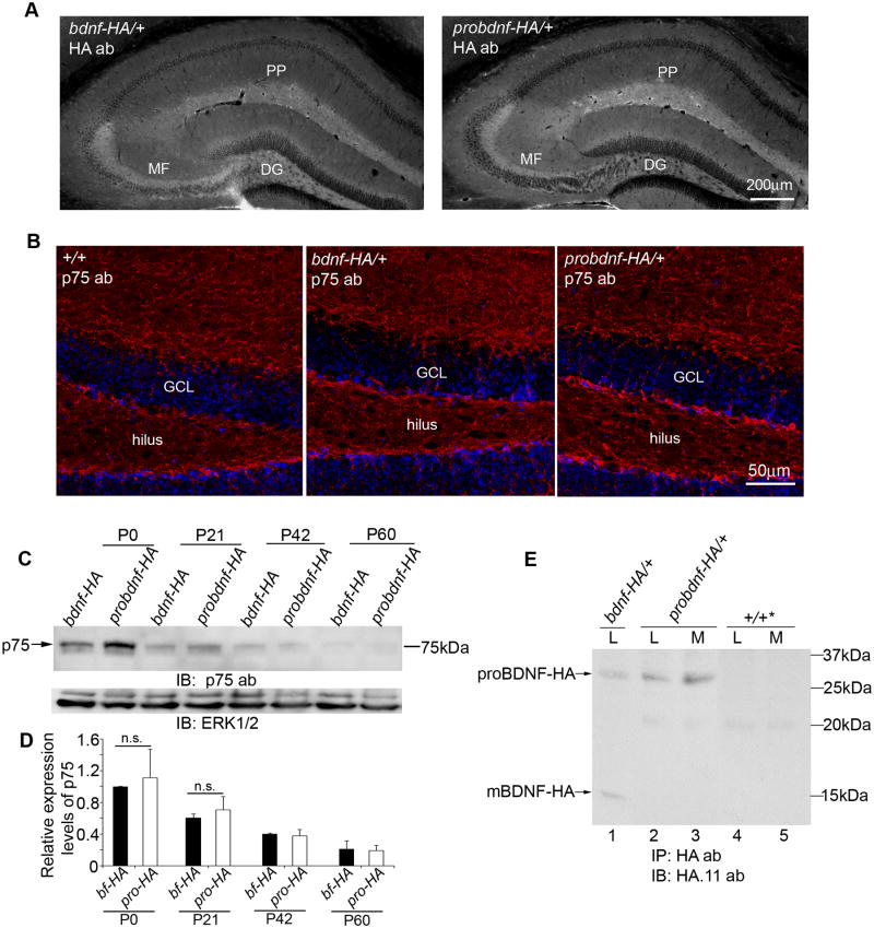
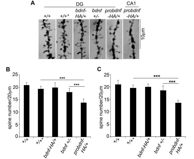
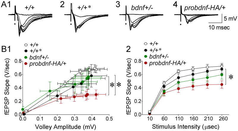
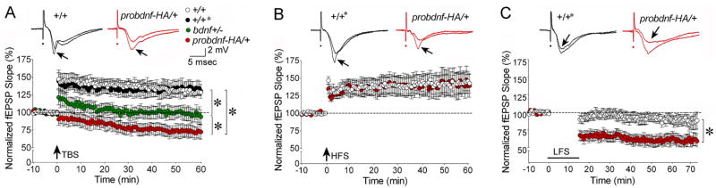
Experience-dependent plasticity shapes postnatal development of neural circuits, but the mechanisms that refine dendritic arbors, remodel spines, and impair synaptic activity are poorly understood. Mature brain-derived neurotrophic factor (BDNF) modulates neuronal morphology and synaptic plasticity, including long-term potentiation (LTP) via TrkB activation. BDNF is initially translated as proBDNF, which binds p75(NTR). In vitro, recombinant proBDNF modulates neuronal structure and alters hippocampal long-term plasticity, but the actions of endogenously expressed proBDNF are unclear. Therefore, we generated a cleavage-resistant probdnf knockin mouse. Our results demonstrate that proBDNF negatively regulates hippocampal dendritic complexity and spine density through p75(NTR). Hippocampal slices from probdnf mice exhibit depressed synaptic transmission, impaired LTP, and enhanced long-term depression (LTD) in area CA1. These results suggest that proBDNF acts in vivo as a biologically active factor that regulates hippocampal structure, synaptic transmission, and plasticity, effects that are distinct from those of mature BDNF.


Figures

Fig.1
Fig.1. Generation of probdnf-HA knock-in mice
A. Developmental expression of proBDNF and mature BDNF. Left: Hippocampi from mice of indicated ages expressing two alleles of bdnf-HA (bdnf-HA/HA) were lysed, and immunoblotting was performed using anti-HA. Immunoblots using anti-ERK1/2 were performed for a loading control. Right: Immunoprecipitation/Western blot analysis of hippocampi or cortices from wildtype (+/+) or bdnf-HA/HA mice of the indicated age. B. Strategy for generating the probdnf-HA knock-in mice is shown schematically. C. Southern blot analysis of ES clones demonstrates that one endogenous bdnf allele was replaced by a probdnf-HA allele. D. Western blot analysis of proBDNF-HA expression in the hippocampi of probdnf-HA/+ mice at 4 months. Hippocampi were lysed and immunoblotting was performed using an anti-HA antibody; anti-ERK1/2 was performed for a loading control. E. Quantitation of total BDNF isoforms (proBDNF + mature BDNF) utilizing ELISA from mice of indicated genotype at 5 months (n=3/genotype). F. Western blot analysis to determine the levels of mature BDNF in probdnf-HA/+ mice, bdnf+/- mice, and wildtype (+/+) mice at 5 months of age. Hippocampi were lysed and immunoblotting was performed using an anti-BDNF antibody; anti-ERK1/2 was performed for a loading control. G. Quantitation of relative levels of mature BDNF from mice of indicated genotype. Hippocampal lysates (5 months) were analyzed by western blotting, and quantitation was performed using ImageJ (*, p<0.01; n.s., not significant; n=3/genotype).
Fig. 2
Fig. 2. Detection of proBDNF-HA and p75NTR, and secretion of proBDNF, from hippocampal neurons of probdnf-HA/+ mice
A. Detection of HA immunoactivity from probdnf-HA/+ and bdnf-HA/+ brains using anti-HA and Cy3-conjugated streptavidin with image-capture in black/white (white corresponds to immunoreactivity). Localization of proBDNF-HA in probdnf-HA/+ mice is comparable to bdnf-HA/+ mice, indicating that proBDNF-HA is transported normally in the mossy fibers in vivo. B. p75NTR localization in dentrate gyrus of probdnf-HA/+, bdnf-HA/+ and wildtype (+/+) mice (7 weeks of age). Immunofluorescence was detected in dendritic processes of granule cells in all genotypes; cellular localization of p75NTR in probdnf-HA/+ mice was comparable to the other genotypes. p75NTR immunoreactivity is noted in red; DAPI is in blue. C. p75NTR expression in postnatal hippocampi from probdnf-HA/+ or bdnf-HA/+ mice of the indicated ages. Lysates were separated by SDS-PAGE, and immunoblotting was performed using anti-p75NTR. Normalization was performed using anti-ERK1/2 detection. D. Quantification of p75NTR expression level in postnatal mouse hippocampi at different ages indicated (3 hippocampi/genotype). n.s., p>0.05. E. Secretion of proBDNF-HA from hippocampal neurons cultured from probdnf-HA/+ and bdnf-HA/+ mice. Neurons were cultured for 7 days in depolarizing conditions. Media was collected, cell lysates prepared and immmunoprecipitated with anti-HA. Mature BDNF-HA was not detected in probdnf-HA/+ lysates and media, but was present in bdnf-HA/+ lysates. No immunoreactivity was detected in wildtype cultures (+/+*), in lysates or media.
Fig. 3
Fig. 3. Altered hippocampal anatomy in probdnf-HA/+ mice
A, B. Sholl analysis of dentate granule cells from P30 (A) or P105 (B) mice. 40 neurons from 4-5 animals were analyzed per genotype. Results are presented as mean ± SEM. Red asterisks indicate significant differences between probdnf-HA/+ mice and controls; black asterisks indicate significant differences between probdnf-HA /+ mice and bdnf+/-mice (p<0.01). C. Representative traces of Golgi-stained granule cells from control mice (+/+), wildtype littermates of probdnf-HA/+ mice (+/+*), bdnf+/- mice, and probdnf-HA/+ mice at P105. D. Sholl analysis of granule cells from P105 mice. To confirm the reduction of dendritic arborization in proBDNF-expressing mice was mediated by p75NTR receptors, Golgi-stained dentate granule neurons from probdnf-HA/+:p75NTR-/-, +/+:p75NTR-/-, p75NTR-/-, and wildtype littermates of probdnf-HA/+ (+/+*) were traced. The deletion of p75NTR rescued the reduction of dendritic arborization in probdnf-HA/+ mice. E. Reduced hippocampal volume in probdnf-HA knock-in mice. Total hippocampal volume was quantitated by Cavalieri analysis of MRI images of brains from 11 month wildtype mice (+/+, n=3), bdnf+/- n=5, bdnf-HA/+ (n=4) and probdnf-HA/+ mice (n=5). Asterisk indicates p<0.01 by Student's t-test. n.s., not significant (p=0.12).
Fig. 4
Fig. 4. proBDNF negatively regulates spine formation
A. Representative images of Golgi-stained dendritic spines of granule neurons (DG) or pyramidal neurons in region CA1 from wildtype mice (+/+), wildtype littermates of probdnf-HA/+ mice (+/+*), bdnf+/-, bdnf-HA/+ and probdnf-HA/+ mice. B, C. Reduction of dendritic spine density in probdnf-HA/+ knock-in mice at 1 month. Brain sections from mice of the indicated genotypes were subjected to Golgi-staining. Dendritic spines of 15 neurons/mouse of 3-4 mice/genotype were counted at 100×. Total spine number along a 20 μm-long dendrite was measured. Probdnf-HA/+ mice had fewer spines compared to bdnf+/- mice in CA1 (B) and the DG (C). Differences were significant (Student's t-tests, ***, p<0.0001).
Fig. 5
Fig. 5. Basal transmission is adversely affected in probdnf-HA/+ mice
A. FEPSPs elicited at several stimulus strengths are superimposed. Representative responses to stimulation are shown for +/+ mice (wildtype littermates of bdnf+/- mice (14 slices, 7 mice), +/+* (wildtype littermates of probdnf-HA/+ mice; 21 slices, 13 mice), bdnf+/- mice (10 slices, 5 mice) and probdnf-HA/+ mice (22 slices, 15 mice). B. 1. FEPSP slope is plotted in relation to fiber volley amplitude. Probdnf-HA/+ mice were more severely affected than +/+* mice (ANCOVA, p=0.04) and bdnf+/- mice (p=0.02). 2. FEPSP slope is plotted in relation to stimulus strength, reflected by the duration of a constant current (100 μA) stimulus. Probdnf-HA/+ mice were more severely affected than littermate controls (+/+*; two-way RMANOVA, p<0.05). Probdnf-HA/+ mice were not significantly different from bdnf+/- mice (two-way RMANOVA, p>0.05) but were significantly impaired relative to bdnf+/- mice at the maximal stimulus.
Fig. 6
Fig. 6. TBS-LTP is impaired, HFS-LTP is unaffected, and LTD is enhanced in probdnf-HA/+ mice
A. Top: Representative fEPSPs, pre- (no arrow) and 60 min post- (arrow) TBS. Bottom: TBS-LTP was most impaired in probdnf-HA/+ mice. There were significant differences (p<0.05, large asterisks) except for +/+ vs. +/+* genotypes. B. Top: Representative fEPSPs, pre- (no arrow) and 60 min post- (arrow) HFS. Bottom: Differences in HFS-LTP between probdnf-HA/+ mice (red), and wildtype littermates (+/+*; white) were not significant. C. Top: Representative fEPSPs, pre- (no arrow) and 60 min post- (arrow) LFS. Bottom: LTD was greater in probdnf-HA/+ mice compared to +/+* (p<0.05).

References

    1. Bath KG, Voss HU, Jing D, Anderson S, Hempstead B, Lee FS, Dyke JP, Ballon DJ. Quantitative intact specimen magnetic resonance microscopy at 3.0 T. Magn Reson Imaging. 2009;27:672–80. - PMC - PubMed
    1. Bramham CR. Local protein synthesis, actin dynamics, and LTP consolidation. Curr Opin Neurobiol. 2008;18:524–31. - PubMed
    1. Chen G, Kolbeck R, Barde YA, Bonhoeffer T, Kossel A. Relative contribution of endogenous neurotrophins in hippocampal long-term potentiation. J Neurosci. 1999;19:7983–90. - PMC - PubMed
    1. Chen ZY, Jing D, Bath KG, Ieraci A, Kha T, Siao CJ, Herrera DG, Toth M, Yang C, McEwen BS, Hempstead BL, Lee FS. Genetic variant BDNF (Val66Met) polymorphism alters anxiety-related behavior. Science. 2006;314:140–3. - PMC - PubMed
    1. Conner JM, Lauterborn JC, Yan Q, Gall CM, Varon S. Distribution of brain-derived neurotrophic factor (BDNF) protein and mRNA in the normal adult rat CNS: evidence for anterograde axonal transport. J Neurosci. 1997;17:2295–313. - PMC - PubMed

Publication types

MeSH terms