Review
. 2014 Jun;18(6):279-92.
doi: 10.1016/j.tics.2014.03.002. Epub 2014 Apr 18.

Neural mechanisms of motivated forgetting

Affiliations
Review

Neural mechanisms of motivated forgetting

Michael C Anderson et al. Trends Cogn Sci. 2014 Jun.

Abstract

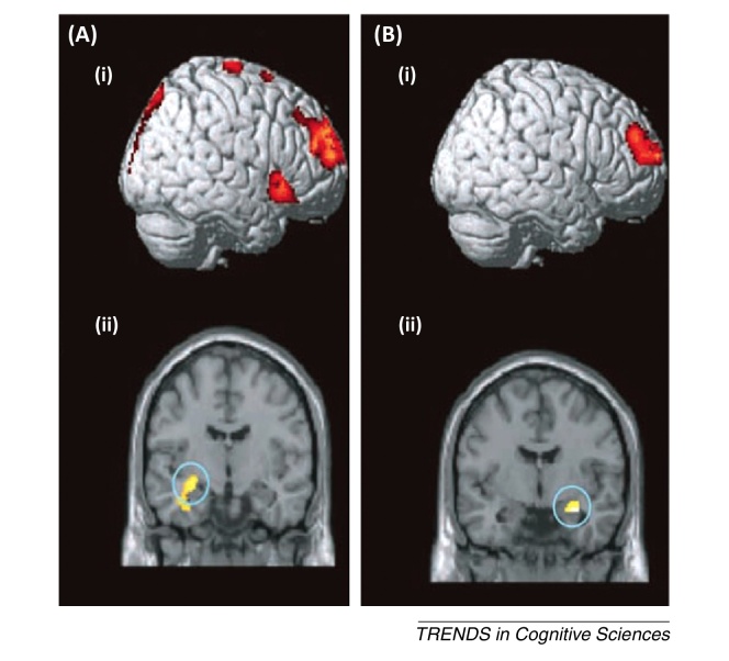
Not all memories are equally welcome in awareness. People limit the time they spend thinking about unpleasant experiences, a process that begins during encoding, but that continues when cues later remind someone of the memory. Here, we review the emerging behavioural and neuroimaging evidence that suppressing awareness of an unwelcome memory, at encoding or retrieval, is achieved by inhibitory control processes mediated by the lateral prefrontal cortex. These mechanisms interact with neural structures that represent experiences in memory, disrupting traces that support retention. Thus, mechanisms engaged to regulate momentary awareness introduce lasting biases in which experiences remain accessible. We argue that theories of forgetting that neglect the motivated control of awareness omit a powerful force shaping the retention of our past.


Figures

Figure 1
Figure 1
Neural correlates of directed forgetting (DF). (A) An activation map of a recent item-method directed forgetting functional (f)MRI study . Red areas illustrate significant voxels (P <0.005) indicating greater activity for to-be-forgotten items that are actually forgotten compared with to-be-remembered items that are remembered. (B,C) The results of a multimodal list-method DF experiment . (B) Forget instructions were associated with increased blood oxygenation level-dependent (BOLD) signal in the left dorsolateral prefrontal cortex (DLPFC) and reduced alpha/beta long-range synchrony [11–18 Hz (i)], which were negatively correlated on a single trial level (ii). (C) Stimulating the DLPFC with repetitive transcranial magnetic stimulation (rTMS; 1 Hz) selectively increased list-1 forgetting, without affecting list-2 enhancement. Adapted, with permission, from (B,C). Abbreviation: EEG, electroencephalogram.
Figure 2
Figure 2
Conditions that trigger inhibitory modulation of the hippocampus during retrieval suppression. (A) Direct suppression and thought substitution involve distinct networks that both cause forgetting, but that have differing effects on the hippocampus . Direct suppression involves suppressing episodic retrieval to prevent or override recollection of an unwanted memory (depicted by angled lines), whereas thought substitution involves engaging retrieval to recall a substitute thought in response to a reminder. Direct suppression (upper row) engages right dorsolateral prefrontal cortex (DLPFC) and ventrolateral prefrontal cortex (VLPFC), with the former reducing hippocampal activity (established by effective connectivity analyses). By contrast, thought substitution (lower row) engages a left dominant VLPFC region that does not reduce hippocampal activity (and in fact, predicts increased hippocampal activity [88]). (B) Measuring intrusions on no-think trials using a trial-by-trial intrusion scale (left, upper portion; ratings of 2 or 3 indicate an intrusion of the to-be-suppressed memory) reveals intrusions that decline with repeated suppressions (left lower) . Strikingly, although suppression reduces hippocampal activity overall (right panel, top left subpanel; green bar, think; red bar, no-think), this modulation is driven strongly by trials on which intrusions occur (right panel, top right subpanel, red bar, intrusions; orange bar, non-intrusions). Hippocampal downregulation (pre-trial - no-think activation, z-normalized) predicts later memory deficits (baseline - no-think, z-normalized) during intrusions, but not during nonintrusions (right panel, bottom). Abbreviation: fMRI, functional MRI.
Figure 3
Figure 3
How suppressing retrieval reduces the unconscious influence of unwanted memories, via neocortical inhibition . (A) Adaptation of the think/no-think (TNT) procedure (67). After learning word–object associations, participants either repeatedly retrieved (think) or suppressed (no-think) objects, using direct suppression . On the final test, participants viewed objects distorted by noise that were gradually revealed, and participants indicated when they could identify the distorted object. (B) Suppressing retrieval activated the right dorsolateral prefrontal cortex (DLPFC) (i), and reduced activity in fusiform gyrus (ii) (effective connectivity analyses established that the former modulated the latter). (C) Behavioural and neural aftereffects of suppressing visual memories. All objects showed repetition priming (speeded identification time), relative to novel objects, but this was reduced for suppressed objects (i). Similarly, all studied objects showed neural priming (reduced neural activity) in fusiform gyrus and the lateral occipital complex, relative to novel objects, but this was partially reversed for suppressed objects (ii). Negative coupling between DLPFC and fusiform gyrus predicted the magnitude of the reversal in neural priming on the final perceptual identification test (iii). Abbreviations: DCM, Dynamic Causal Modelling; MGF, middle frontal gyrus; ROI, region of interest.
Figure I
Figure I
The item and list-methods for studying directed forgetting, along with the typical pattern of findings (for real examples, see and , respectively).
Figure I
Figure I
Brain-imaging data from two patients with dissociative amnesia . Patients 1 (A) and 2 (B) viewed images of faces and decided whether they recognised them from their life. Images were either strangers (novel), faces they knew, from outside the window of amnesia (identifiable faces) or faces they knew from within the amnesic window (unidentifiable faces). (Ai) and (Bi) depict brain areas that are more active for unidentifiable faces than for identifiable faces (right dorsolateral prefrontal cortex). (Aii) and (Bii) depict brain areas that are less active for unidentifiable faces (hippocampus).

References

    1. Baddeley A. Psychology Press; 2009. Memory.
    1. Bjork R.A. Retrieval inhibition as an adaptive mechanism in human memory. In: Roediger H.L., Craik F.I.M., editors. Varieties of Memory and Consciousness: Essays in Honour of Endel Tulving. Erlbaum; 1989. pp. 309–330.
    1. Sahakyan L. List-method directed forgetting in cognitive and clinical research: a theoretical and methodological review. In: Ross B.H., editor. Volume 59. Elsevier; 2013. pp. 131–189. (Psychology of Learning and Motivation).
    1. Muther W.S. Erasure of partitioning in short-term memory. Psychon. Sci. 1965;3:429–430.
    1. Geiselman R.E.R. Disrupted retrieval in directed forgetting: a link with posthypnotic amnesia. J. Exp. Psychol. Gen. 1983;112:58–72. - PubMed

Publication types