Review
. 2014 Aug;1839(8):719-27.
doi: 10.1016/j.bbagrm.2014.04.011. Epub 2014 Apr 18.

Structural biology-based insights into combinatorial readout and crosstalk among epigenetic marks

Affiliations
Review

Structural biology-based insights into combinatorial readout and crosstalk among epigenetic marks

Jiamu Du et al. Biochim Biophys Acta. 2014 Aug.

Abstract

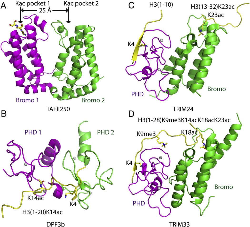
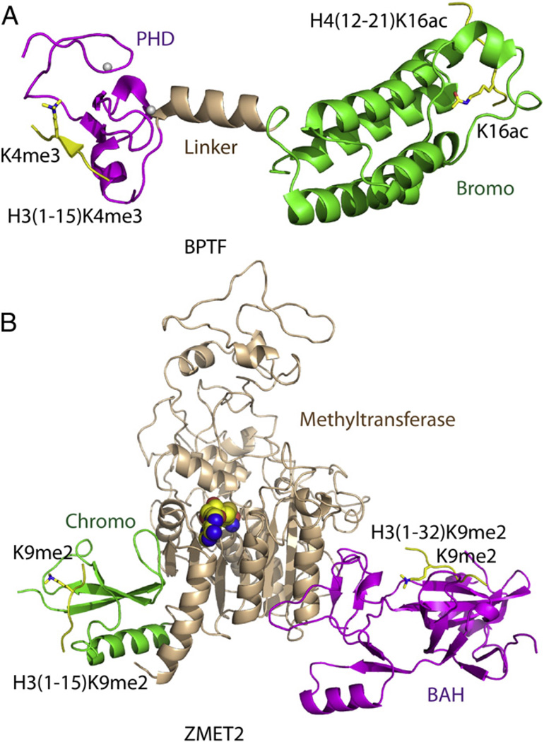
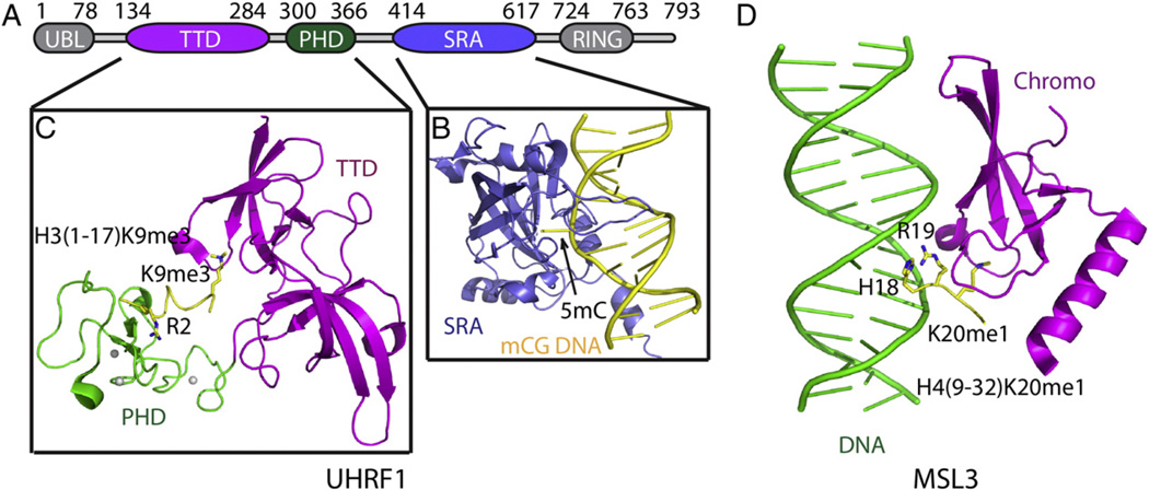
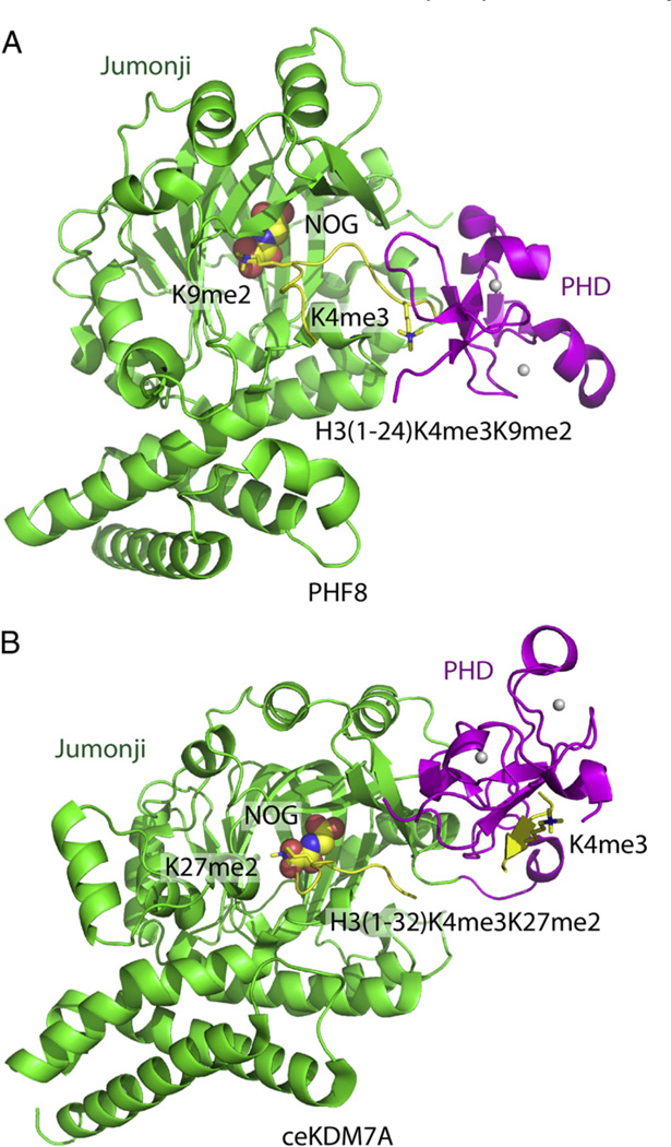
Epigenetic mechanisms control gene regulation by writing, reading and erasing specific epigenetic marks. Within the context of multi-disciplinary approaches applied to investigate epigenetic regulation in diverse systems, structural biology techniques have provided insights at the molecular level of key interactions between upstream regulators and downstream effectors. The early structural efforts focused on studies at the single domain-single mark level have been rapidly extended to research at the multiple domain-multiple mark level, thereby providing additional insights into connections within the complicated epigenetic regulatory network. This review focuses on recent results from structural studies on combinatorial readout and crosstalk among epigenetic marks. It starts with an overview of multiple readout of histone marks associated with both single and dual histone tails, as well as the potential crosstalk between them. Next, this review further expands on the simultaneous readout by epigenetic modules of histone and DNA marks, thereby establishing connections between histone lysine methylation and DNA methylation at the nucleosomal level. Finally, the review discusses the role of pre-existing epigenetic marks in directing the writing/erasing of certain epigenetic marks. This article is part of a Special Issue entitled: Molecular mechanisms of histone modification function.

Keywords: Combinatorial readout; DNA methylation; Epigenetic regulation; Histone modification.


Figures

Fig. 1
Fig. 1
Structural basis for multivalent readout of histone marks from a single histone tail. (A) Ribbon representation of the crystal structure of TAFII250 double bromodomain (PDB code: 1EQF) with bromodomain 1 colored in magenta and bromodomain 2 in green. The N-terminus of a symmetry related protein inserts into the acetyllysine (Kac) binding pocket of bromodomain 1. The distance between two Kac binding pockets of the two bromodomains is 25 Å. (B) Ribbon representation of the solution NMR structure of DPF3b double PHD fingers in complex with H3(1–20)K14ac peptide (PDB code: 2KWJ)with PHD1 finger colored in magenta, the PHD2 finger in green, and the bound peptide in yellow. The zinc ions are shown as silver balls. The specific residues recognized on the H3 peptide, including K4 and K14ac, are highlighted in stick representations. (C) Ribbon-representation model of TRIM24 PHD-Bromo cassette simultaneously recognizing unmodified H3K4 and H3K23ac following superposition of the crystal structures of TRIM24 PHD-Bromo-H3(1–10) complex (PDB code: 3O37) and TRIM24 PHD-Bromo-H3(13–32)K23ac complex (PDB code: 3O34). The PHD finger, bromodomain and bound peptides are colored in magenta, green and yellow, respectively. The unmodified H3K4 and H3K23ac are highlighted in stick representations. (D) Ribbon representation of the crystal structure of TRIM33 PHD-Bromo cassette in complex with H3(1–28)K9me3/K14ac/K18ac/K23ac peptide (PDB code: 3U5O and 3U5P) with the PHD finger, bromodomain, and bound peptide colored in magenta, green and yellow, respectively. The unmodified H3K4, H3K9me3 and H3K18ac, which are specifically recognized, are highlighted in stick representations.
Fig. 2
Fig. 2
Structural basis for multivalent readout of multiple histone tails. (A) Ribbon-representation of a model of the BPTF PHD-Bromo cassette simultaneously recognizing H3K4me3 and H4K16ac following superposition of the crystal structures of BPTF PHDBromo-H3(1–15)K4me3 complex (PDB code: 2F6J) and BPTF PHD-Bromo-H4(12–21) K16ac complex (PDB code: 3QZS). The PHD finger, the bromodomain, the linker region and the bound peptides are colored in magenta, green, wheat and yellow, respectively. TheH3K4me3 and H4K16ac are highlighted in stick representations. (B) Ribbon representation of a model of ZMET2 simultaneously recognizing two H3K9me2 peptides with its BAH domain and chromodomain by superposition of the crystal structures of ZMET2-H3(1–15)K9me2 complex (PDB code: 4FT2) and ZMET2-H3(1–32)K9me2 complex (PDB code: 4FT4). The BAH domain, the methyltransferase, the chromodomain and the bound peptides are colored in magenta, wheat, green and yellow, respectively. The two H3K9me2 marks are highlighted in stick representations.
Fig. 3
Fig. 3
Structural basis for multivalent readout of histone and DNA marks. (A) The domain architecture of multiple domain protein UHRF1, which can recognize both histone marks and hemimethylated CpG DNA. (B) Ribbon representation of the structure of the UHRF1 SRA domain in complex with a hemimethylated CpG DNAwith the SRA domain (PDB code: 3CLZ) and the DNA colored in slate and yellow, respectively. The 5mC base is flipped out of the DNA helix and indicated by an arrow. (C) Ribbon representation of the crystal structure of UHRF1 tandem Tudor domain (TTD)-PHD cassette in complex with the H3(1–17)K9me3 peptide (PDB code: 4GY5). The TTD domain, the PHD finger domain and the bound peptide are colored inmagenta, green and yellow, respectively. The specifically recognized unmodified H3R2 and H3K9me3 residues are highlighted in stickmodel. (D) Ribbon representation of the crystal structure of MSL3 chromodomain in complex with a DNA duplex and H4(9–32)K20me1 peptide (PDB code: 3OA6). The chromodomain, DNA and peptide are colored inmagenta, green and yellow, respectively. The peptide residues H18 and R19, which interact with DNA, and H20me1, which is specifically recognized by the chromodomain, are highlighted in stick representations.
Fig. 4
Fig. 4
Structural basis for recognition by histone modification-directed histone modification enzyme. (A) Ribbon representation of the crystal structure of PHF8 in complex with H3(1–24)K4me3/K9me2 peptide (PDB code: 3KV4). The PHD finger, Jumonji domain and the bound peptide are colored in magenta, green and yellow, respectively. The NOG cofactor is shown in a space-filling representation. The H3K4m3mark, which is specifically recognized by the PHD finger, and the H3K9me2 mark, which specifically inserts into the active site of the Jumonji domain, are highlighted in stick representations. The PHF8 enzyme shows a bent conformation upon K4me3 binding into the PHD pocket, with the distance between the PHD pocket and Jumonji active site short enough to allow the H3K9me2 mark to simultaneously insert into the Jumonji active site. (B) Ribbon representation of the crystal structure of ceKDM7A in complex with H3(1–32)K4me3/K27me2 peptide (PDB code: 3N9P). The PHD finger, Jumonji domain and the bound peptide are colored in magenta, green and yellow, respectively. The NOG cofactor is shown in a space filling representation. The H3K4me3 mark, which is specifically recognized by the PHD finger, and the H3K27me2 mark, which specifically inserts into the active site of the Jumonji domain, are highlighted in stick representation. The ceKDM7A enzyme shows an extended conformation upon K4me3 binding into the PHD pocket, with the distance between the PHD pocket and Jumonji active site being too long to allow simultaneous recognition of H3K9me2, but should allow simultaneous recognition of H3K27me2 mark by inserting it into the Jumonji active site.

References

    1. Luger K, Mader AW, Richmond RK, Sargent DF, Richmond TJ. Crystal structure of the nucleosome core particle at 2.8 A resolution. Nature. 1997;389:251–260. - PubMed
    1. Jenuwein T, Allis CD. Translating the histone code. Science. 2001;293:1074–1080. - PubMed
    1. Law JA, Jacobsen SE. Establishing, maintaining and modifying DNA methylation patterns in plants and animals. Nat. Rev. Genet. 2010;11:204–220. - PMC - PubMed
    1. Liu Y, Zhang X, Blumenthal RM, Cheng X. A common mode of recognition for methylated CpG. Trends Biochem. Sci. 2013;38:177–183. - PMC - PubMed
    1. Feng S, Jacobsen SE, Reik W. Epigenetic reprogramming in plant and animal development. Science. 2010;330:622–627. - PMC - PubMed

Publication types

MeSH terms

LinkOut - more resources