. 2014 Jun;13(6):822-31.
doi: 10.1128/EC.00341-13. Epub 2014 Apr 18.

Trypanosoma cruzi bromodomain factor 3 binds acetylated α-tubulin and concentrates in the flagellum during metacyclogenesis

Affiliations

Trypanosoma cruzi bromodomain factor 3 binds acetylated α-tubulin and concentrates in the flagellum during metacyclogenesis

Victoria Lucia Alonso et al. Eukaryot Cell. 2014 Jun.

Abstract

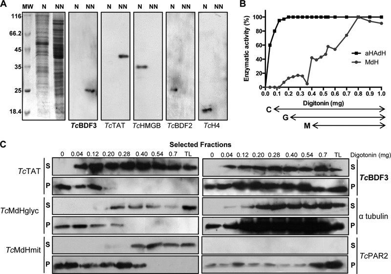
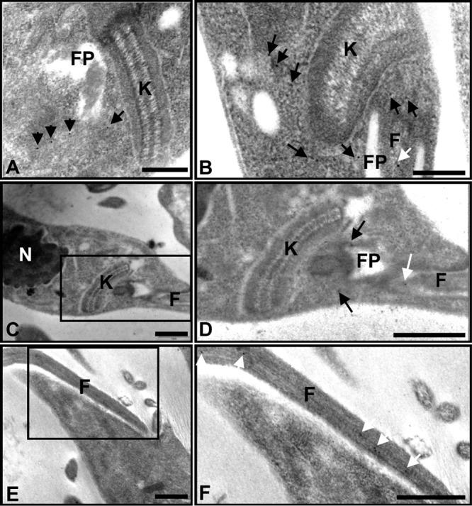
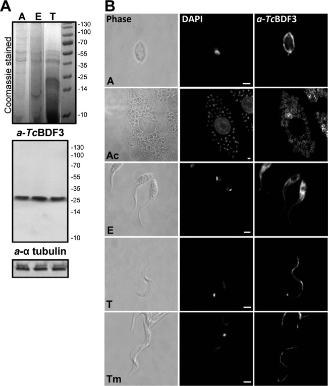
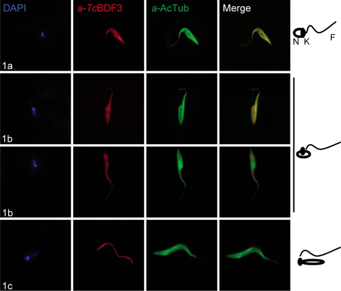
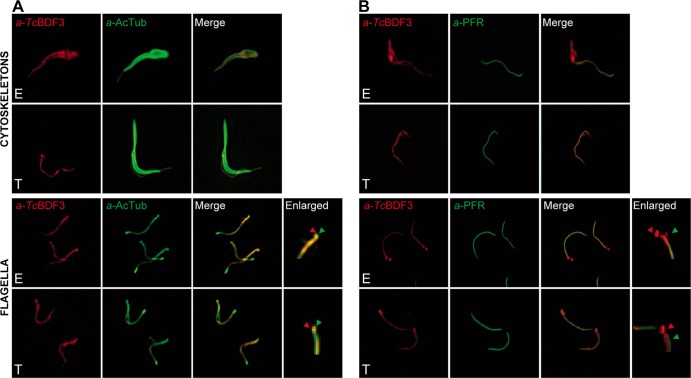
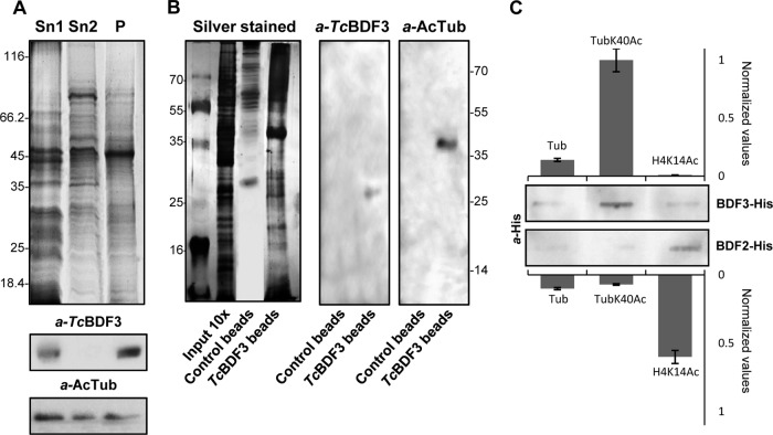
Bromodomains are highly conserved acetyl-lysine binding domains found mainly in proteins associated with chromatin and nuclear acetyltransferases. The Trypanosoma cruzi genome encodes at least four bromodomain factors (TcBDFs). We describe here bromodomain factor 3 (TcBDF3), a bromodomain-containing protein localized in the cytoplasm. TcBDF3 cytolocalization was determined, using purified antibodies, by Western blot and immunofluorescence analyses in all life cycle stages of T. cruzi. In epimastigotes and amastigotes, it was detected in the cytoplasm, the flagellum, and the flagellar pocket, and in trypomastigotes only in the flagellum. Subcellular localization of TcBDF3 was also determined by digitonin extraction, ultrastructural immunocytochemistry, and expression of TcBDF3 fused to cyan fluorescent protein (CFP). Tubulin can acquire different posttranslational modifications, which modulate microtubule functions. Acetylated α-tubulin has been found in the axonemes of flagella and cilia, as well as in the subpellicular microtubules of trypanosomatids. TcBDF3 and acetylated α-tubulin partially colocalized in isolated cytoskeletons and flagella from T. cruzi epimastigotes and trypomastigotes. Interaction between the two proteins was confirmed by coimmunoprecipitation and far-Western blot assays with synthetic acetylated α-tubulin peptides and recombinant TcBDF3.


Figures

FIG 1
FIG 1
TcBDF3 is a cytoplasmic bromodomain-containing protein in epimastigotes. (A) Nuclear (N) and nonnuclear (NN) protein extracts (30 μg per well) were subjected to Western blot analysis using rabbit anti-TcBDF3 antibodies, anti-TcTAT (cytosolic), anti-TcHMGB (nuclear), anti-TcBDF2 (nuclear), and anti-T. cruzi H4 histone (TcH4) (nuclear). On the left is the Coomassie-stained gel. (B) Enzymatic activity of αHAdH and MdH in epimastigotes treated with increasing concentrations of digitonin. Activities were measured and normalized to the protein concentration in the extracts. The arrows below the graph indicate the digitonin concentrations at which cytosolic (C), glycosomal (G), and mitochondrial (M) proteins are released. (C) Equal volumes of selected soluble (S) and insoluble (P) fractions obtained at different digitonin amounts (0 to 0.7 mg) were subjected to Western blot analysis using anti-TcBDF3 antibodies (TcBDF3) and known markers for different organelles. The antibodies used were anti-TcTAT (cytosolic), anti-TcMdHglyc (glycosomal), anti-TcMdHmit (mitochondrial), anti-TcPAR2 (flagellar), and anti-α-tubulin (α-tubulin) (cytoskeletal). TL, total lysate.
FIG 2
FIG 2
TcBDF3 is localized at the cytoplasm, the flagellum, and the flagellar pocket of epimastigotes. (A to C and E) Immunoelectron microscopy of TcBDF3 in T. cruzi epimastigotes using purified rabbit anti-TcBDF3 antibodies. The nucleus (N), kinetoplast (K), flagellar pocket (FP), and flagellum (F) are indicated. Gold particles are indicated with black and white arrows. The white arrows indicate flagellar labeling. (D) Enlarged image of the boxed area in panel C. (F) Enlarged image of the boxed area in panel E. Bars = 1 μm.
FIG 3
FIG 3
TcBDF3 is expressed throughout the T. cruzi life cycle in vitro. (A) Western blot analysis using purified rabbit anti-TcBDF3 antibodies (a-TcBDF3) and mouse anti-α-tubulin (a-α-tubulin) as a loading control. A, amastigote total protein extracts; E, epimastigote total protein extracts; T, trypomastigote total protein extracts (30 μg per well). (B) Immunofluorescence assay using purified anti-TcBDF3 and parasites at different stages of the T. cruzi life cycle. A, free amastigote; Ac, amastigotes inside a Vero cell; E, epimastigote; T, trypomastigote from infected Vero cells; Tm, metacyclic trypomastigote obtained in vitro. Anti-rabbit IgG conjugated to fluorescein was used as a secondary antibody. Nuclei and kinetoplasts were labeled with DAPI. Bars = 2 μm.
FIG 4
FIG 4
TcBDF3 changes its location during in vitro metacyclogenesis. Immunofluorescence assays used purified rabbit anti-TcBDF3 (α-TcBDF3) and mouse monoclonal anti-acetylated α-tubulin (α-AcTub) antibodies in intermediate stages 1a, 1b, and 1c, as defined by Ferreira et al. (51). On the right are schematic diagrams of the positions of the flagellum (F), nucleus (N), and kinetoplast (K) in the three selected intermediate differentiation stages. Anti-rabbit IgG conjugated to fluorescein (green) and anti-mouse IgG conjugated to rhodamine (red) were used as secondary antibodies. Nuclei and kinetoplasts were labeled with DAPI (blue).
FIG 5
FIG 5
TcBDF3 is detected in the cytoskeletons and flagella of epimastigotes (E) and only in the flagella of metacyclic trypomastigotes (T). Immunofluorescence assays used purified rabbit anti-TcBDF3 (α-TcBDF3) and mouse monoclonal anti-acetylated α-tubulin (α-AcTub) (A) and mouse anti-PAR2 (α-PFR) (B) antibodies on isolated cytoskeletons and flagella of epimastigotes and metacyclic trypomastigotes. Anti-rabbit IgG conjugated to fluorescein (green) and anti-mouse IgG conjugated to rhodamine (red) were used as secondary antibodies. The last right lanes are enlarged images of the detergent-resistant structures that correspond to MTs attached to basal bodies and forming the flagellar pocket. The green arrowheads indicate TcBDF3 localization, and the red arrowheads indicate acetylated α-tubulin (A) and PAR2 (B) localization in these structures.
FIG 6
FIG 6
TcBDF3 interacts with acetylated α-tubulin. (A) Coomassie-stained SDS-PAGE and Western blot analyses of epimastigote protein extracts enriched in cytoskeletal and flagellar proteins. Sn1, soluble protein extracts; Sn2, soluble cytoskeletal and flagellar protein extracts; P, insoluble cytoskeletal and flagellar protein extracts (50 μg per well). Rabbit anti-TcBDF3 antibodies (α-TcBDF3) and mouse anti-acetylated α-tubulin antibodies (α-AcTub) were used. (B) Coimmunoprecipitation assay using purified anti-TcBDF3 antibodies covalently coupled to magnetic beads (TcBDF3 beads). Magnetic beads coupled to IgGs (purified from antisera of nonimmunized rabbits) were used as a negative control (Control beads). On the left is a silver-stained SDS-PAGE gel of total cytoskeletal extracts and the elutions obtained after the immunoprecipitation experiment. On the right is a Western blot analysis of the eluted proteins after coimmunoprecipitation using purified rabbit anti-TcBDF3 antibodies (α-TcBDF3) and mouse monoclonal anti-acetylated α-tubulin antibodies (α-AcTub). (C) Slot far-Western blot assay. Acetylated tubulin (TubK40Ac), nonacetylated tubulin (Tub), and acetylated histone H4 (H4K14Ac) peptides were blotted onto a nitrocellulose membrane and incubated with His-tagged recombinant TcBDF3 (BDF3-His) or TcBDF2 (BDF2-His). Bound recombinant proteins were detected with anti-histidine antibodies (α-His). Signals were quantified by densitometry and normalized using the interaction with acetylated α-tubulin peptide as a reference (assigned the arbitrary value of 1). The bars and error bars indicate means ± standard deviations (SD) from the results of three independent experiments.

References

    1. Contreras VT, Araujo-Jorge TC, Bonaldo MC, Thomaz N, Barbosa HS, de Meirelles MN, Goldenberg S. 1988. Biological aspects of the Dm 28c clone of Trypanosoma cruzi after metacyclogenesis in chemically defined media. Mem. Inst. Oswaldo Cruz 83:123–133. 10.1590/S0074-02761988000100016 - DOI - PubMed
    1. Hill KL. 2010. Parasites in motion: flagellum-driven cell motility in African trypanosomes. Curr. Opin. Microbiol. 13:459–465. 10.1016/j.mib.2010.05.015 - DOI - PMC - PubMed
    1. Field MC, Carrington M. 2009. The trypanosome flagellar pocket. Nat. Rev. Microbiol. 7:775–786. 10.1038/nrmicro2221 - DOI - PubMed
    1. Hammond JW, Cai D, Verhey KJ. 2008. Tubulin modifications and their cellular functions. Curr. Opin. Cell Biol. 20:71–76. 10.1016/j.ceb.2007.11.010 - DOI - PMC - PubMed
    1. Verhey KJ, Gaertig J. 2007. The tubulin code. Cell Cycle 6:2152–2160. 10.4161/cc.6.17.4633 - DOI - PubMed

Publication types

MeSH terms

LinkOut - more resources