. 2014 Apr 18;9(4):e92836.
doi: 10.1371/journal.pone.0092836. eCollection 2014.

Hematopoietic overexpression of FOG1 does not affect B-cells but reduces the number of circulating eosinophils

Affiliations

Hematopoietic overexpression of FOG1 does not affect B-cells but reduces the number of circulating eosinophils

Camille Du Roure et al. PLoS One. .

Abstract

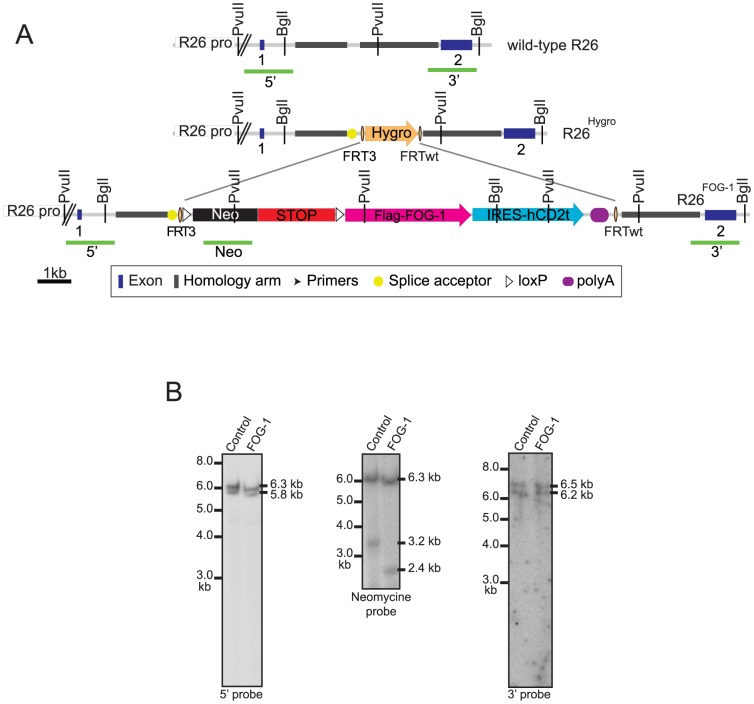
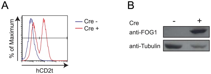
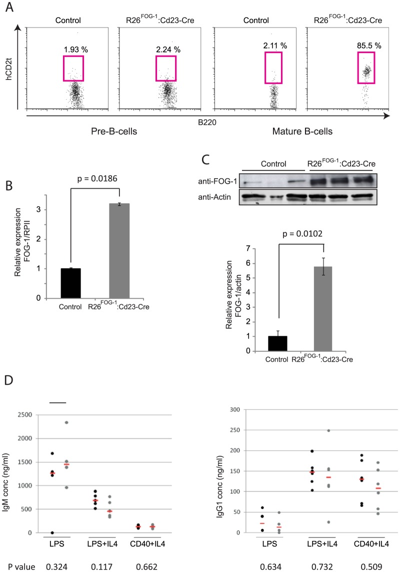
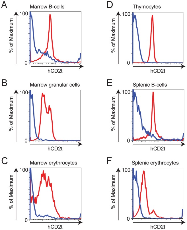
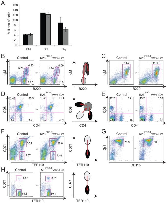
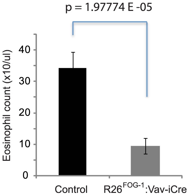
We have identified expression of the gene encoding the transcriptional coactivator FOG-1 (Friend of GATA-1; Zfpm1, Zinc finger protein multitype 1) in B lymphocytes. We found that FOG-1 expression is directly or indirectly dependent on the B cell-specific coactivator OBF-1 and that it is modulated during B cell development: expression is observed in early but not in late stages of B cell development. To directly test in vivo the role of FOG-1 in B lymphocytes, we developed a novel embryonic stem cell recombination system. For this, we combined homologous recombination with the FLP recombinase activity to rapidly generate embryonic stem cell lines carrying a Cre-inducible transgene at the Rosa26 locus. Using this system, we successfully generated transgenic mice where FOG-1 is conditionally overexpressed in mature B-cells or in the entire hematopoietic system. While overexpression of FOG-1 in B cells did not significantly affect B cell development or function, we found that enforced expression of FOG-1 throughout all hematopoietic lineages led to a reduction in the number of circulating eosinophils, confirming and extending to mammals the known function of FOG-1 in this lineage.


Conflict of interest statement

Competing Interests: Thierry Doll and Matthias Müller are employed by Novartis Institutes for Biomedical Research. Camille Du Roure is employed by Actelion. There are no patents, products in development or marketed products to declare. This does not alter the authors' adherence to all the PLOS ONE policies on sharing data and materials.

Figures

Figure 1
Figure 1. FOG-1 is expressed in a regulated manner during B-cell development.
(A) Gene expression level of FOG-1 determined by Affymetrix microarray in wild-type or OBF-1−/− Abl Pro-B-cells . (B) Reverse transcriptase (RT)-PCR analysis of FOG-1 mRNA level in wild-type or OBF-1−/− Abl Pro-B-cells. GAPDH mRNA amount was used as a control. +, − refer to cDNA synthesis reactions performed in the presence or absence of reverse transcriptase (RT), respectively. (C) mRNA expression level of FOG-1 in bone marrow B-cells (B220+) and in red blood cells (TER 119+) determined by semi-quantitative RT-PCR. Actin mRNA amount was used as a reference. (D) mRNA expression level of FOG-1 during B-cell development determined by semi-quantitative RT-PCR. The analysis was performed on primary Pro-B, large Pre-B, small Pre-B, immature-B and splenic mature-B cells. Actin mRNA amount was used as a reference. (E) mRNA expression level of FOG-1 measured by real-time RT-PCR during the B-cell development. The analysis was performed on primary B-cells as in (D). RNA Polymerase II mRNA was used as a reference. (F) mRNA expression level of FOG-1 in B-cell lines representing different stages of B-cell development, determined by RT-PCR. From early to late stages, the analysis was performed on the five following cell lines: wild-type Abelson (Abl+/+), B3, A20J, X63 and J558. GAPDH mRNA was used as a control.
Figure 2
Figure 2. Strategy for the rapid generation of conditionally overexpressing ES cells.
Schematic representation of the wild-type Rosa26 (R26) locus, the pre-targeted R26Hygro and the recombined R26 alleles. The Rosa26 locus was first pre-targeted by homologous recombination with a cassette containing a splice acceptor and a hygromycin B resistance gene flanked by a FRT3 site in 5′ and a FRTwt site in 3′ to generate the pre-targeted R26Hygro allele (step 1). The hygromycin B cassette is then replaced by the cassette of interest via the FRT sites using transient expression of the FLP recombinase (step 2). Our cassette of interest contains a loxP-Neo-STOP-loxP cassette for neomycin selection of ES cells and conditional expression of the transgene, and an IRES-hCD2t sequence to monitor expressing cells. Notably, Cre-mediated recombination will excise the neomycin gene from the targeted allele in the transgenic mice. Neo: neomycin resistance gene. R26 pro: promoter of the R26 gene.
Figure 3
Figure 3. Correct recombination at the pre-targeted R26Hygro allele by RMCE.
A. Schematic representation of the wild-type R26, pre-targeted R26Hygro and recombined R26FOG-1 alleles. In the FOG-1 cassette, the neomycin resistance gene (Neo, black) and the STOP sequence (red) are flanked by LoxP sites (white triangles) to allow conditional expression. The cDNA encoding FlagFOG-1 (pink) followed by an IRES sequence and the sequence coding for hCD2t (pale blue) were inserted downstream. A polyA signal was placed at the 3′ end of the hCD2t coding sequence (blue oval). The probes (green bars) used for Southern blot analysis are shown. B. Correct insertion of the vectors was confirmed by Southern blotting. To test the 5′ boundary, PvuII-digested ES cell genomic DNA was hybridized with the radioactively labeled 5′ probe to detect the wild-type (5.8 kb) and the targeted (6.3 kb) bands (Left panel). To verify single-copy insertion, PvuII-digested DNA was hybridized with a radioactively labeled internal probe to detect the 3.2 kb or 2.4 kb targeted bands in control or FOG-1 clones, respectively, in addition to the 6.3 kb targeted band (Middle panel). To test the 3′ boundary, BglI-digested DNA was hybridized with the radioactively labeled 3′ probe to detect the 6.5 kb wild-type and the 6.2 kb targeted bands (Right panel).
Figure 4
Figure 4. Cre-dependent expression of hCD2t and FOG-1 in recombined ES cells.
R26FOG-1 ES cells were electroporated with a Cre expressing vector. Cre-induced expression of hCD2t and FOG-1 was detected by flow cytometry (A) and western blot analysis (B), respectively.
Figure 5
Figure 5. Overexpression of FOG-1 in mature B-cells.
A. In R26FOG-1:Cd23-Cre mice, hCD2t expression is restricted to mature B-cells. Cell surface expression of hCD2t was analyzed by flow cytometry in Pre-B-cells (B220+, CD25+) and in mature B-cells (B220+, IgMhigh) of control (R26FOG-1) and R26FOG-1:Cd23-Cre mice. Representative results of at least 3 independent experiments are shown. B. FOG-1 mRNA is increased 3-fold in R26FOG-1:Cd23-Cre mature B-cells. RNA extracted from 3 control (R26FOG-1) and 3 R26FOG-1:Cd23-Cre mice was reverse transcribed and subjected to quantitative PCR to detect FOG-1 and RNA Polymerase II (RPII, for normalization) transcripts. Standard error of the mean is shown. C. FOG-1 protein is up-regulated ca. 6-fold in mature B-cells derived from R26FOG-1:Cd23-Cre mice. FOG-1 and actin proteins were detected by western blotting in mature B-cells derived from 3 control (R26FOG-1) and 3 R26FOG-1:Cd23-Cre mice (upper panel). The band intensities were quantified by LiCor Odyssey scanning and normalized to expression of actin (lower panel). Standard error of the mean is shown. D. FOG-1-overexpressing mature B-cells respond normally to in vitro stimulation. Splenic resting mature B-cells isolated from 3 to 6 control (R26FOG-1, black dots) or R26FOG-1:Cd23-Cre mice (grey dots) were activated in vitro by LPS, LPS+IL4 or anti-CD40+IL4 for 4 days. Titers of IgM (left panel) or IgG1 (right panel) in the culture supernatants were determined by ELISA; means are shown (red bar) as well as the corresponding p values at the bottom.
Figure 6
Figure 6. hCD2t is expressed in all hematopoietic cells of R26FOG-1:Vav-iCre mice.
A–F. Flow cytometry analysis of hCD2t expression in R26FOG-1:Vav-iCre (red line) and control (R26FOG-1, blue line) mice. A. Bone marrow B-lymphocytes (B220+ cells). B. Bone marrow granular cells (based on Forward and Side Scatters). C. Bone marrow erythrocytes (TER119+ cells). D. Thymocytes (CD4+, CD8+ cells). E. Splenic B-lymphocytes (B220+ cells). F. Splenic erythrocytes (TER119+ cells). Data for one representative animal of each genotype are shown (n = 5).
Figure 7
Figure 7. Normal B-cell, T-cell and granular cell populations in R26FOG-1:Vav-iCre mice.
A. Cells of the bone marrow (BM), spleen (Spl) and thymus (Thy) of R26FOG-1 (black bars) and R26FOG-1:Vav-iCre (grey bars) mice were enumerated. Standard error of the mean is shown. B. Bone marrow cells were stained with anti-B220 and anti-IgM antibodies to analyze B-cell development. C. Splenocytes were stained with anti-B220 and anti-IgM antibodies to identify B-cells. D. Thymocytes were stained with anti-CD4 and anti-CD8 antibodies to analyze T-cell development. E. Splenocytes were stained with anti-CD4 and anti-CD8 antibodies to identify mature T-cells. F. Bone marrow cells were stained with anti-TER119 and anti-CD71 antibodies to analyze erythropoiesis. G. Bone marrow cells were stained with anti-Gr1 and anti-CD11b antibodies to identify Gr1+ CD11b+ myeloid cells. H. Splenocytes were stained with anti-TER119 and anti-CD71 antibodies to analyze splenic erythropoiesis. Cells were analyzed by flow cytometry in R26FOG-1 (control) and R26FOG-1:Vav-iCre animals; data for one representative animal are shown (n = 5 for each genotype). Percentages of the populations are shown next to the gates. A diagram representing the developmental pathway of the different lineages from pale (progenitors) to dark grey (differentiated cells) is shown next to the pseudo-dotplots B, D, F and H.
Figure 8
Figure 8. Altered eosinophil numbers in R26FOG-1:Vav-iCre mice.
Reduction of circulating eosinophils. The numbers of eosinophils obtained in full blood count analysis of 8 control (R26FOG-1) and 8 R26FOG-1:Vav-iCre including those presented in Table 1 mice were averaged. Standard error of the mean is shown.

References

    1. Matthias P, Rolink AG (2005) Transcriptional networks in developing and mature B cells. Nature reviews Immunology 5: 497–508. - PubMed
    1. Orkin SH, Zon LI (2008) Hematopoiesis: an evolving paradigm for stem cell biology. Cell 132: 631–644. - PMC - PubMed
    1. Cantor AB, Orkin SH (2005) Coregulation of GATA factors by the Friend of GATA (FOG) family of multitype zinc finger proteins. Seminars in cell & developmental biology 16: 117–128. - PubMed
    1. Tsang AP, Fujiwara Y, Hom DB, Orkin SH (1998) Failure of megakaryopoiesis and arrested erythropoiesis in mice lacking the GATA-1 transcriptional cofactor FOG. Genes Dev 12: 1176–1188. - PMC - PubMed
    1. Fox AH, Liew C, Holmes M, Kowalski K, Mackay J, et al. (1999) Transcriptional cofactors of the FOG family interact with GATA proteins by means of multiple zinc fingers. The EMBO journal 18: 2812–2822. - PMC - PubMed

Publication types

LinkOut - more resources