Review
. 2014 Jul:31:124-31.
doi: 10.1016/j.semcdb.2014.04.019. Epub 2014 Apr 18.

Cellular foundations of mammary tubulogenesis

Affiliations
Review

Cellular foundations of mammary tubulogenesis

Robert J Huebner et al. Semin Cell Dev Biol. 2014 Jul.

Abstract

The mammary gland is composed of a highly branched network of epithelial tubes, embedded within a complex stroma. The mammary epithelium originates during embryonic development from an epidermal placode. However, the majority of ductal elongation and bifurcation occurs postnatally, in response to steroid hormone and growth factor receptor signaling. The process of pubertal branching morphogenesis involves both elongation of the primary ducts across the length of the fat pad and a wave of secondary branching that elaborates the ductal network. Recent studies have revealed that mammary epithelial morphogenesis is accomplished by transitions between simple and stratified organization. During active morphogenesis, the epithelium is stratified, highly proliferative, has few intercellular junctions, and exhibits incomplete apico-basal polarity. In this review, we discuss recent advances in our understanding of the relationship between epithelial architecture, epithelial polarity, and ductal elongation.

Keywords: Apico-basal polarity; Branching morphogenesis; Breast cancer; Collective cell migration; Mammary gland; Tubulogenesis.


Figures

Fig. 1
Fig. 1
(A) Mammary development in the mouse begins at embryonic day 12 when an epidermal placode invades the mammary stroma. The stratified epithelium then elongates and hollows to form a rudimentary bilayered duct that remains essentially quiescent until puberty. Modified from Hogg et al, 1983. (B) Mammary ducts are elaborated during puberty to form the functional ductal network. Pubertal development initiates during postnatal week 3 with the formation of terminal end buds (TEBs), which are a stratified epithelium at the ductal tips. Over the next 7 weeks, primary ducts elongate across the mammary fat pad, and secondary branching completes the ductal tree.
Fig. 2
Fig. 2
(A) Mammary epithelium consists of a rudimentary ductal network prior to puberty. (A’) Duct from a 2-week-old mouse stained for F-actin (red) and nuclei (green). All mammary ducts are bilayered before pubertal development. Modified from Huebner, Lechler, and Ewald, 2014. (B) TEBs form at ductal tips at the onset of puberty. (B’) TEB from a 4-week-old mouse stained for F-actin (red) and nuclei (green). TEBs consist of a stratified epithelium with more than two cell layers. Modified from Huebner, Lechler and Ewald, 2014. (C) TEBs function as the elongation front of the mammary epithelium during pubertal development. (C’) TEB stained for the basolateral polarity marker ß-catenin (red), the tight junction protein zona occludins 1 (ZO-1), (green) and nuclei (blue). TEBs have incomplete apico-basal polarity with ß-catenin present on nearly all cellular membranes and tight junctions present primarily at the apical-most membrane facing the luminal space. Aside from the primary luminal space, TEBs also have microlumens, highlighted with arrows, which are positive for tight junctions. Modified from Ewald et al, 2008.
Fig. 3
Fig. 3
(A) Migration of single cells involves front-rear polarization and actin-based protrusions at the leading edge. (B) A possible mechanism for ductal elongation is migration of a subset of polarized protrusive leader cells. (C) Organoid bud stained for F-actin (green) and nuclei (red). (C’) Zoomed image of the elongation front of the bud shown in C. Actin-based protrusions are noticeably absent from the leading edge of elongating mammary branches. Modified from Ewald et al, 2008. (D) Electron micrograph of an elongating mammary bud. (D’) Leading edge of the bud shown in D. Elongating mammary branches have a smooth leading edge when viewed at the ultrastructural level. Modified from Ewald et al, 2012.
Fig. 4
Fig. 4
(A) Model of mammary duct and TEB structure. Ducts are a bilayered epithelium with apical luminal epithelial cells (gray) and basal myoepithelial cells (brown). Luminal cells have tight junctions (green) at the apical-most point of cell-cell contact. Desmosomes (blue) are cell adhesion complexes that connect luminal cells to each other and to the myoepithelial cell layer. TEBs are composed of a low-polarity internal cell population (light blue) that is bounded by the basal myoepithelial layer and apical luminal cells. Internal cells have a diminished number of desmosomes and loose cell-cell contacts by electron microscopy. Tight junctions are absent from internal cells except at microlumens. Mammary ducts and TEBs are surrounded by basement membrane (red). (B) Schematic showing the apico-basal polarity of luminal epithelial cells. The polarity protein par-3 (orange) is localized to the apical membrane and scribble (red) is localized to the basolateral membrane. ZO-1 (green) marks the tight junction, which partitions the apical and basolateral membranes. (C) Model of the cellular mechanism of mammary duct stratification. Stratification is initiated by vertical proliferation of apically localized luminal epithelial cells, and the vertical divisions result in asymmetric localization of tight junctions and polarity loss for internal cells. Stratification is non-clonal, as multiple vertical divisions are observed within a single organoid. Low-polarity internal cells are proliferative, further driving stratification. Modified from Huebner, Lechler, and Ewald, 2014. (D) Vertical divisions resulted in asymmetric localization of par-3 (orange) ZO-1 (green) and scribble (red) in the apical mother cell and the internal daughter cell. This asymmetry directly results in polarity loss for the internal daughter cell. The cell fate marker numb (blue) is asymmetrically localized to the basal daughter cell during vertical divisions. Modified from Huebner, Lechler and Ewald, 2014.
Fig. 5
Fig. 5
(A) Schematic showing each stage of mammary branching morphogenesis. Prior to development, ducts are bilayered with apical luminal epithelial cells (gray) and basal myoepithelial cells (brown), and the entire duct is surrounded by basement membrane (red). The epithelium then stratifies, forming a low-polarity internal cell population (light blue). Low-polarity TEBs then elongate the epithelial ducts, and finally the epithelium polarizes back to a bilayer. (B) Still images from a movie of an organoid mosaically labeled with cytosolic GFP to visualize single cells within the epithelium. Cells are labeled green, the membranes are red, and the dashed line shows the elongation front. Cells were protrusive and migratory within the organoids. The arrow highlights a cell that moves to the basal surface and then back into the body of the elongating branch. Modified from Ewald et al, 2012.
Fig. 6
Fig. 6
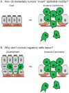
(A) Cartoon depiction of epithelial cell dissemination past the basement membrane. (B) Schematic questioning why normal motile cells within the epithelium do not invade past the basement membrane.

References

    1. Nelson WJ. Remodeling epithelial cell organization: transitions between front-rear and apical-Basal polarity. Cold Spring Harb Perspect Biol. 2009;1:a000513. - PMC - PubMed
    1. O’Brien LE, Zegers MM, Mostov KE. Opinion: Building epithelial architecture: insights from three-dimensional culture models. Nat Rev Mol Cell Biol. 2002;3:531–537. - PubMed
    1. Bilder D. Epithelial polarity and proliferation control: links from the Drosophila neoplastic tumor suppressors. Genes Dev. 2004;18:1909–1925. - PubMed
    1. McNally S, Martin F. Molecular regulators of pubertal mammary gland development. Ann Med. 2011;43:212–234. - PubMed
    1. Veltmaat JM, Mailleux AA, Thiery JP, Bellusci S. Mouse embryonic mammogenesis as a model for the molecular regulation of pattern formation. Differentiation. 2003;71:1–17. - PubMed

Publication types

LinkOut - more resources