. 2014 May;20(5):503-10.
doi: 10.1038/nm.3521. Epub 2014 Apr 20.

Essential role for autophagy in the maintenance of immunological memory against influenza infection

Affiliations

Essential role for autophagy in the maintenance of immunological memory against influenza infection

Min Chen et al. Nat Med. 2014 May.

Abstract

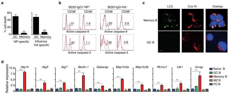
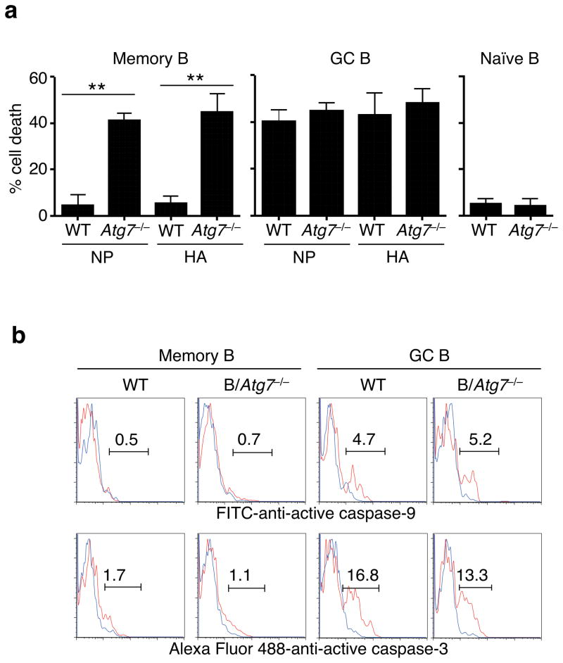
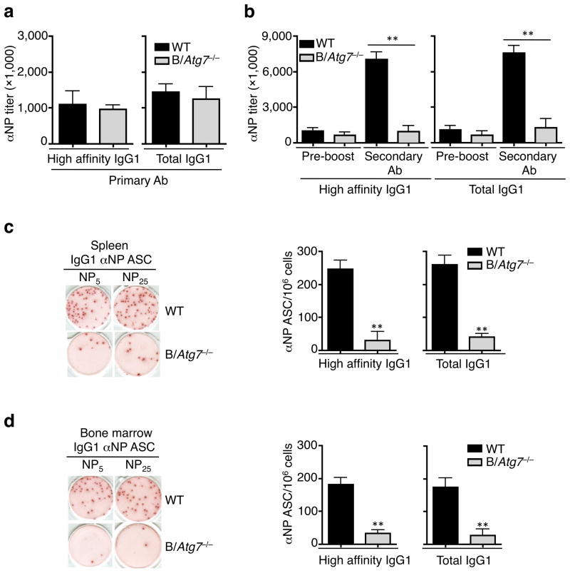
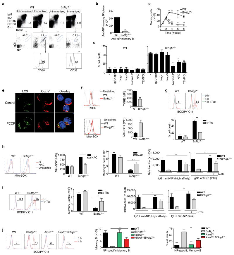
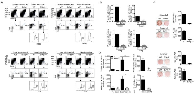
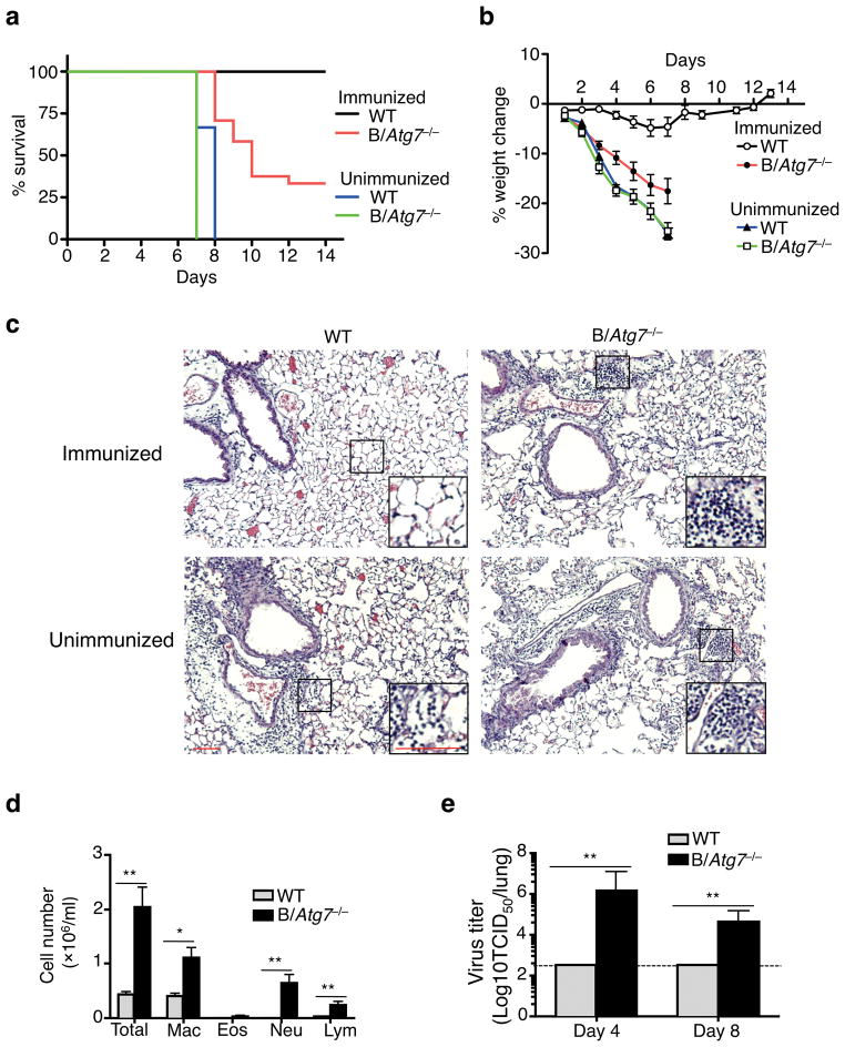
Vaccination has been the most widely used strategy to protect against viral infections for centuries. However, the molecular mechanisms governing the long-term persistence of immunological memory in response to vaccines remain unclear. Here we show that autophagy has a critical role in the maintenance of memory B cells that protect against influenza virus infection. Memory B cells displayed elevated levels of basal autophagy with increased expression of genes that regulate autophagy initiation or autophagosome maturation. Mice with B cell-specific deletion of Atg7 (B/Atg7(-/-) mice) showed normal primary antibody responses after immunization against influenza but failed to generate protective secondary antibody responses when challenged with influenza viruses, resulting in high viral loads, widespread lung destruction and increased fatality. Our results suggest that autophagy is essential for the survival of virus-specific memory B cells in mice and the maintenance of protective antibody responses required to combat infections.


Figures

Figure 1
Figure 1. Decreased spontaneous cell death and caspase signaling but constitutive autophagy in memory B cells
(a) Percentages of cell loss of NP- or HA-specific memory or GC B cells after in vitro culture. Experiments were performed four times in triplicates using cells from a pool of 15 mice. **P<0.01. (b) Intracellular staining of active caspase-3 or caspase-9 in NP- or HA-specific memory or GC B cells with (red line) or without (blue line) in vitro culture. Data are representative of three experiments. (c) Immunocytochemistry for LC3 and CoxIV staining in NP-specific memory and GC B cells. Data are representative of three experiments. Scale bar: 5 μm. (d) Real-time RT-PCR analyses of autophagy-related genes in naïve mature, germinal center (GC), memory, marginal zone (MZ) and follicular (FO) B cells. Experiments were performed three times in triplicates using cells from a pool of 15 mice. Data in this figure are presented as mean ± SEM. **P<0.01 (determined by two-tailed Student’s t-test).
Figure 2
Figure 2. Increased cell death in autophagy-deficient memory B cells
(a) Cell death of NP- or HA-specific memory B cells, GC B cells or naïve B cells from B/Atg7−/− mice or CD19-cre mice as wild type (WT) controls after in vitro culture. Experiments were performed three times in triplicates using cells from a pool of 20 mice. Data are presented as mean ± SEM. Comparison to WT control: **P<0.01 (determined by two-tailed Student’s t-test). (b) Intracellular staining of active caspase-3 or caspase-9 in NP-specific memory or GC B cells with (red line) or without (blue line) in vitro culture. Data are representative of four independent experiments.
Figure 3
Figure 3. Normal primary but defective secondary antibody responses in B/Atg7−/− mice
(a) Titers of IgG1 anti-NP antibodies detected with NP5 (high-affinity) or NP25 (total) two weeks after immunization (n=10 mice per group). Data are presented as mean ± SEM representative of three independent experiments. Differences between WT and B/Atg7−/− mice are statistically insignificant. (b) Titers of high-affinity and total IgG1 anti-NP antibodies in the sera of immunized mice before boosting or 5 days after antigen boosting. **P<0.01 (n=10 per group in each experiment). Data are presented as mean ± SEM and are representative of three independent experiments. **P<0.01 (determined by two-tailed Student’s t-test). (c, d) ELISPOT for NP-specific IgG1 ASC in the spleen (c) and bone marrow (d) of mice immunized as in (a). A representative image is also shown. Data are presented as mean ± SEM (n=10 per group in each experiment) that are representative of three independent experiments. **P<0.01 (determined by two-tailed Student’s t-test).
Figure 4
Figure 4. Loss of memory B cells in the absence of Atg7
(a, b) DUMPB220+IgG1+CD38+NP+ memory B cells in the spleen of B/Atg7−/− mice or WT controls eight weeks after immunization. Unimmunized mice were used as negative controls (a). Total NP-specific memory B cells in the spleen are presented in (b). **P<0.01 (n=10 mice/group). Data are representative of five independent experiments. (c) The total numbers of NP-specific memory B cells in the spleen of B/Atg7−/− mice or WT controls at indicated days after NP-KLH immunization. Data are representative of two independent experiments. **P<0.01 (n=5 mice/group at each time point). (d) Cell death of NP-specific memory B cells in the presence of different inhibitors after in vitro culture. **P<0.01 (n=3). Data are representative of three experiments. (e) Immunocytochemistry staining for LC3 and CoxIV in NP-specific memory B cells with or without treatment with FCCP. Data are representative of two independent experiments. LC3 punctates/cell: untreated, 10.8 ± 2.4; FCCP, 28.4 ± 4.3. **P<0.01 (n=10). Scale bar: 5 μm. (f) Staining of NP-specific memory B cells with TMRE or Mito-SOX. Mean fluorescence intensity (MFI) is also presented. Data are representative of three independent experiments. **P<0.01 (n=3). (g) Staining with BODIPY in memory B cells after culture for 4 h in the presence or absence of α-Toc. Percentages of positive staining without α-Toc treatment (red line) are shown. Percentages of memory B cell death were also quantified. **P<0.01 (n=6). (h) Mito-SOX staining and total counts of memory B cells from the spleen of NP-KLH-immunized mice injected with NAC or PBS. Induction of NP-specific secondary antibodies was also measured in parallel experiments. **P<0.01 (n=6 mice/group). (i) BODIPY staining and total counts of memory B cells from the spleen of NP-KLH-immunized mice treated with α-Toc. Induction of NP-specific secondary antibodies was also measured in parallel experiments. **P<0.01 (n=6 mice/group). (j) BODIPY staining of NP-specific memory B cells after in vitro culture for 0 or 4 h. Total numbers of memory B cells in the spleen of Alox5−/−B/Atg7−/− and control mice two months after NP-KLH immunization were quantitated. Memory B cell death was measured as in (d). **P<0.01 (n=6). Data in this figure are presented as mean ± SEM (P value are determined by two-tailed Student’s t-test).
Figure 5
Figure 5. Defective memory B cell responses to influenza infection in B/Atg7−/− mice
(a, b) HA-specific memory B cells in the spleen and lung of B/Atg7−/− mice or wild type controls two months after intranasal immunization with influenza virus at a sublethal dose of 7.5 TCID50. Unimmunized mice were used as negative controls (a). The total numbers of NP-specific memory B cells in the spleen and lung are presented (b). Data are representative of three independent experiments. Quantitative analyses are presented as mean ± SEM. **P<0.01 (6 mice/group). (c) Mice immunized as in (a) and re-challenged with influenza virus 2 months later. Anti-HA IgG or IgA in the sera or BAL fluids was quantitated 6 days after viral challenge. Antibody titers in the sera before the virus re-challenge are also shown. Data are representative of three independent experiments. Quantitative analyses are presented as mean ± SEM. **P<0.01 (n=10/group). (d) Anti-HA antibody secreting cells (ASC) in the spleen and lung of mice in (c) were quantitated by ELISPOT. Data are presented as mean ± SEM from three independent experiments. **P<0.01 (n=10/group).
Figure 6
Figure 6. B/Atg7−/− mice are defective in mounting protective immunity against influenza virus
(a, b) B/Atg7−/− mice and WT controls with or without prior influenza immunization were challenged with a lethal dose of influenza virus two months later. The percentage of survival or loss of body weight was quantitated (WT immunized, n=15; B/Atg7−/− immunized, n=21; WT unimmunized, n=10; B/Atg7−/− unimmunized, n=10). B/Atg7−/− versus WT immunized group, P<0.0001 (determined by log-rank test). Data are representative of three independent experiments. (c) Mice with or without influenza virus immunization were infected as in (a) and used for histochemistry analyses of the lung 6 days after virus re-challenge (n=3 per group). Scale bar: 100 μm. The area indicated by the small square was amplified and shown on the lower right corner. Scale bar: 100 μm. (d) Quantitation of BAL fluid cells in the lung from mice with prior immunization and re-challenged as in (a). *P<0.05, **P<0.01 (n=6 per group) (determined by two-tailed Student’s t-test). Data are representative of two independent experiments. (e) Quantitation of virus load in the lung from mice immunized and re-challenged as in (a). Data are representative of two independent experiments. **P<0.01 (day 4: n=5 per group; day 8: n=11 for B/Atg7−/−, n=5 for WT) (determined by two-tailed Student’s t-test).

References

    1. Pulendran B, Ahmed R. Immunological mechanisms of vaccination. Nat Immunol. 2011;12:509–517. - PMC - PubMed
    1. Lenardo M, et al. Mature T lymphocyte apoptosis--immune regulation in a dynamic and unpredictable antigenic environment. Annu Rev Immunol. 1999;17:221–253. - PubMed
    1. Strasser A, Jost PJ, Nagata S. The many roles of FAS receptor signaling in the immune system. Immunity. 2009;30:180–192. - PMC - PubMed
    1. Krammer PH, Arnold R, Lavrik IN. Life and death in peripheral T cells. Nat Rev Immunol. 2007;7:532–542. - PubMed
    1. Kalia V, Sarkar S, Gourley TS, Rouse BT, Ahmed R. Differentiation of memory B and T cells. Curr Opin Immunol. 2006;18:255–264. - PubMed

Publication types