. 2014 Jul;80(13):3868-78.
doi: 10.1128/AEM.00759-14. Epub 2014 Apr 18.

Reconstruction of mreB expression in Staphylococcus aureus via a collection of new integrative plasmids

Affiliations

Reconstruction of mreB expression in Staphylococcus aureus via a collection of new integrative plasmids

Ana Yepes et al. Appl Environ Microbiol. 2014 Jul.

Abstract

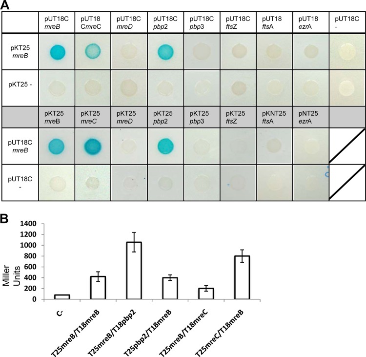
Protein localization has been traditionally explored in unicellular organisms, whose ease of genetic manipulation facilitates molecular characterization. The two rod-shaped bacterial models Escherichia coli and Bacillus subtilis have been prominently used for this purpose and have displaced other bacteria whose challenges for genetic manipulation have complicated any study of cell biology. Among these bacteria is the spherical pathogenic bacterium Staphylococcus aureus. In this report, we present a new molecular toolbox that facilitates gene deletion in staphylococci in a 1-step recombination process and additional vectors that facilitate the insertion of diverse reporter fusions into newly identified neutral loci of the S. aureus chromosome. Insertion of the reporters does not add any antibiotic resistance genes to the chromosomes of the resultant strains, thereby making them amenable for further genetic manipulations. We used this toolbox to reconstitute the expression of mreB in S. aureus, a gene that encodes an actin-like cytoskeletal protein which is absent in coccal cells and is presumably lost during the course of speciation. We observed that in S. aureus, MreB is organized in discrete structures in association with the membrane, leading to an unusual redistribution of the cell wall material. The production of MreB also caused cell enlargement, but it did not revert staphylococcal shape. We present interactions of MreB with key staphylococcal cell wall-related proteins. This work facilitates the use S. aureus as a model system in exploring diverse aspects of cellular microbiology.


Figures

FIG 1
FIG 1
Molecular toolbox for genomic manipulation of S. aureus. (A) Scheme of plasmid pSceI. The I-SceI recognition sequence is adjacent to the I-SceI gene. The bla gene encodes a β-lactamase (ampicillin resistance), and the ermC gene encodes an rRNA methylase (erythromycin resistance). The bgaB gene encodes a β-galactosidase. (B) Scheme of plasmids pAmy (i) and pLac (ii). (C) Representation of the diverse possibilities of cargoes that are inserted into plasmids pAmy and pLac. (i) Transcriptional fusions to the fluorescent proteins GFP and Mars (green and red fluorescence, respectively). (ii) Promoters inducible by IPTG and xylose for protein expression. (iii) His6 and SNAP tags for protein detection.
FIG 2
FIG 2
Physiological assays of the wild-type strain and the amy- and lac-defective mutants. (A) Growth analysis of wild-type (WT) strain Newman and the Δamy and Δlac strains. The OD540 was monitored for 96 h in TSB cultures grown at 37°C. (B) Competition assays of mixed populations (1:1 ratio) of WT and Δamy::YFP strains (Δamy) and of WT and Δlac::YFP strains (Δlac). Mixed communities were incubated at an initial OD540 of 0.05. Samples were taken over 96 h, and CFU were counted by using a dissection scope equipped with a fluorescence excitation detection system. (C) Biofilm formation assay of cultures grown in 24-well titer plates. Biofilms were stained with crystal violet (1%) and quantified by spectrophotometry analysis (n = 3) (error bars represent standard deviations). (D) Hemolytic activity of the different strains. Cultures grown overnight were spotted onto TSB agar containing 5% sheep blood and incubated at 37°C during 48 h. Quantification of hemolytic activity was performed by measuring the OD405 of a solution of 2% erythrocytes previously incubated with the strains' supernatants (n = 3) (error bars represent standard deviations). (E) Fluorescence microscopy pictures used to monitor the cell sizes of the different strains. Cultures were grown until they reached the exponential phase. The membrane of the cell was stained with Nile red (false colored in red), and the DNA was stained with Hoechst 3342 (false colored in blue). Quantification of the cell diameter was performed with Leica Application software (see Materials and Methods).
FIG 3
FIG 3
S. aureus cells expressing mreB of B. subtilis. (A) Western blot analysis detecting the presence of the MreB protein in cell extracts by using polyclonal antibodies against MreB of B. subtilis. WT is wild-type strain Newman. mreB+ is a Newman strain expressing mreB of B. subtilis under the control of an IPTG-inducible promoter. The positive control (C+) is B. subtilis strain PY79. (B) Comparison of cell sizes of wild-type and mreB+ strains. Cultures were grown until exponential phase was reached, and the cell size was monitored by fluorescence microscopy. The cellular membrane was stained with Nile red (false colored red), and the DNA was stained with Hoechst 3342 (false colored blue). Bar, 1 μm. (C) Quantification of the diameters of 500 cells selected from wild-type and mreB+ microscopic fields.
FIG 4
FIG 4
Subcellular distribution of MreB in S. aureus. (A) Fluorescent pictures of S. aureus cells expressing the Mars-MreB translational fusion (false colored red). Shown are Z-stack image series of a single cell expressing mreB. The pictures are sequentially numbered according to their position in the Z-stack. The fluorescence signal was deconvoluted and false-colored red. Bar, 1 μm. A reconstruction of the Z-stack series is presented at the right. In this panel, the fluorescence signals from the 8 planes of the Z-stack are combined into one single plane. Bar, 1 μm. (B) MreB-expressing cells show an altered disposition of cell wall material. Shown are transmission electron microscopy micrographs of sectioned cells from wild-type and MreB-expressing (mreB+) cultures. The unusual reorganization of the cell wall material is observed in discrete regions of the bacterial membrane in MreB-expressing cells, which are labeled with asterisks.
FIG 5
FIG 5
Analysis of protein-protein interactions between MreB and cell wall-related proteins. (A) Bacterial two-hybrid analysis showing interactions between MreB and the protein components of the staphylococcal cell wall elongation machinery (Pbp2, Pbp3, MreC, and MreD) or the cell division protein machinery (FtsZ, FtsA, and EzrA). The MreB-MreB interaction was used as a positive control (pKT25-mreB/pUT18C-mreB). (B) Quantification of positive interactions using β-galactosidase activity. Cells were grown at 30°C for 24 h. Values are the mean results from three independent cultures; error bars represent standard deviations. The Zip/Zip control reached 3,000 Miller units (results not shown). C−, empty vectors.

Similar articles

Cited by

References

    1. Henning UL. 1975. Determination of cell shape in bacteria. Annu. Rev. Microbiol. 29:45–60. 10.1146/annurev.mi.29.100175.000401 - DOI - PubMed
    1. Young KD. 2010. Bacterial shape: two-dimensional questions and possibilities. Annu. Rev. Microbiol. 64:223–240. 10.1146/annurev.micro.112408.134102 - DOI - PMC - PubMed
    1. Errington J, Daniel RA, Scheffers DJ. 2003. Cytokinesis in bacteria. Microbiol. Mol. Biol. Rev. 67:52–65. 10.1128/MMBR.67.1.52-65.2003 - DOI - PMC - PubMed
    1. Pereira PM, Filipe SR, Tomasz A, Pinho MG. 2007. Fluorescence ratio imaging microscopy shows decreased access of vancomycin to cell wall synthetic sites in vancomycin-resistant Staphylococcus aureus. Antimicrob. Agents Chemother. 51:3627–3633. 10.1128/AAC.00431-07 - DOI - PMC - PubMed
    1. Pereira SF, Henriques AO, Pinho MG, de Lencastre H, Tomasz A. 2007. Role of PBP1 in cell division of Staphylococcus aureus. J. Bacteriol. 189:3525–3531. 10.1128/JB.00044-07 - DOI - PMC - PubMed

Publication types

Substances