. 2014 Jul;99(7):1255-63.
doi: 10.3324/haematol.2013.098830. Epub 2014 Apr 18.

Induction of A. fumigatus-specific CD4-positive T cells in patients recovering from invasive aspergillosis

Affiliations

Induction of A. fumigatus-specific CD4-positive T cells in patients recovering from invasive aspergillosis

Hetty Jolink et al. Haematologica. 2014 Jul.

Abstract

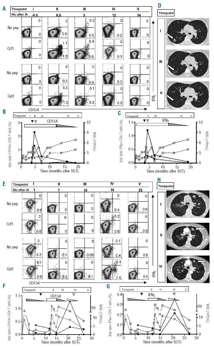
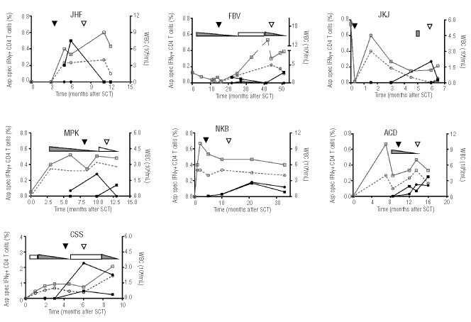
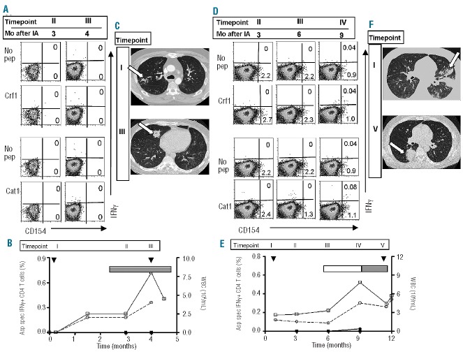
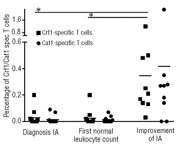
After allogeneic stem cell transplantation patients are at risk of invasive aspergillosis, especially during the period of neutropenia. Recent data suggest that impaired T-cell immune reconstitution after transplantation plays an important role in this increased risk. In this study we investigated whether Aspergillus-specific T cells are involved in the recovery from invasive aspergillosis by analyzing the Aspergillus-specific T-cell response in patients with invasive aspergillosis. In nine patients whose Aspergillus infection improved, we identified Crf1- or Catalase1-specific T cells on the basis of CD154 expression and interferon-γ production following stimulation with overlapping peptides of the A. fumigatus proteins Crf1 and Catalase1. These Aspergillus-specific T cells were induced at the moment of regression of the aspergillus lesions. Crf1- and Catalase1-specific T cells, sorted on the basis of CD154 expression at the peak of the immune response, had a T helper-1 phenotype and recognized a variety of T-cell epitopes. In contrast, in two patients with progressive invasive aspergillosis, no Crf1- or Catalase1-specific T cells were identified. These data indicate that the presence of Aspergillus-specific T cells with a T helper-1 phenotype correlates with the clearance of aspergillus infection.


Figures

Figure 1.
Figure 1.
Aspergillus-specific T-cell responses and imaging studies in patients with improvement of invasive aspergillosis. FACS-analyses imaging studies and kinetics of the Aspergillus-specific T-cell responses of patient MLF (A–D) and patient TPA (E–H). (A and E) FACS analyses were performed on PBMC that were stimulated with Crf1 or Catalase1, and restimulated with Crf1- or Catalase1-pulsed PBMC or with unpulsed PBMC (no pep). T cells were analyzed 5 h after restimulation and FACS-plots are gated on CD4+ T cells. The kinetics of the percentages of Crf1- (■) and Catalase1-specific (•) CD154+ CD4+ T-cells (left y-axis, B and F) and IFNγ+ CD4+ T cells (left y-axis, C and G) and absolute leukocyte (□) and granulocyte counts (○) (right y-axis) are shown before and after diagnosis (▼) and improvement (▽) of aspergillus infection. Use of topical (▭) and systemic (▬) immunosuppression and lowering of the dose (◺,◣) during the study period are depicted in the graphs. Percentages of Aspergillus-specific CD154+ CD4+ T cells were calculated as % CD154+ of CD4+ T cells after restimulation with peptide-pulsed PBMC minus % CD154+ of CD4+ T cells after restimulation with unpulsed PBMC. The same method of calculation was used for IFNγ+ CD4+ T cells. Pulmonary CT scans of patient MLF (D) and patient TPA (H) at different time points after diagnosis show the changes in the size of aspergillus lesions during the course of infection Aspergillus lesions on the CT scans are indicated with an arrow. The timepoints indicated by Roman numerals in the top of the graphs in (B) and (C) and in (F) and (G) correspond to the time points of FACS analyses and CT scans in respectively (A) and (D), and (E) and (H). In figure (A) and (E) the corresponding period in months after invasive aspergillosis (Mo after IA) is given for every timepoint. SCT: stem cell transplantation; WBC: white blood cells.
Figure 2.
Figure 2.
Kinetics of Aspergillus-specific T cells in patients with improvement of aspergillus infection. Percentages of Crf1- (■) and Catalase1-specific (•) IFNγ+ CD4+ T cells (left y-axis) and absolute leukocyte (□) and granulocyte counts (○) (right y-axis) in seven patients before and after diagnosis (▼) and improvement (▽) of aspergillus infection. Use of topical (▭) and systemic (▬) immunosuppression and lowering of the dose (◺,◣) during the study period are depicted in the graphs. Percentages of Aspergillus-specific IFNγ+ CD4+ T cells were calculated as % IFNγ+ of CD4+ T cells after restimulation with peptide-pulsed PBMC minus % IFNγ+ of CD4+ T cells after restimulation with unpulsed PBMC
Figure 3.
Figure 3.
Aspergillus-specific T-cell responses and imaging studies in patients with progressive invasive aspergillosis. FACS analyses, imaging studies and kinetics of the Aspergillus-specific T-cell responses of patient MST (A–C) and ESF (D–F). (A and D) FACS analyses were performed on PBMC after restimulation with Crf1- or Catalase1-pulsed PBMC or with unpulsed PBMC (no pep). T cells were analyzed 5 h after restimulation and FACS-plots are gated on CD4+ T cells. (B and E) Percentages of Crf1- (■) and Catalase1-specific (•) IFNγ+ CD4+ T cells (left y-axis) and absolute leukocyte (□) and granulocyte counts (○) (right y-axis) after diagnosis (▼) of aspergillus infection. Use of topical (▭) and systemic (▬) immunosuppression during the study period is depicted in the graphs. Percentages of Aspergillus-specific IFNγ+ CD4+ T cells were calculated as % IFNγ+ of CD4+ T cells after restimulation with peptide-pulsed PBMC minus % IFNγ+ of CD4+ T cells after restimulation with unpulsed PBMC. Pulmonary CT scans of patient MST (C) and ESF (F) show progressive disease with new nodular lesions at later time points. Aspergillus lesions on the CT scans are indicated with an arrow. The time points indicated by Roman numerals in the top of the graphs in (B) and (E) correspond to the time points of FACS analyses and CT scans in, respectively, (A) and (C), and (D) and (F). In (A) and (D) the corresponding period in months after invasive aspergillosis (Mo after IA) is given for every time point.
Figure 4.
Figure 4.
Increases of Crf1- and Catalase1-specific T cells at the time of improvement of aspergillus infection. Percentages of Crf1- (■) and Catalase1-specific (•) IFNγ+ CD4+ T cells around diagnosis of aspergillus infection (11 patients), at the first time point with normal leukocyte counts (11 patients) and at the time of improvement of aspergillus infection (9 patients). *Significant difference between compared groups (P<0.01, two-tailed Student T test) Horizontal lines indicate the mean percentage of Crf1- or Catalase1-specific T cells among all analyzed patients at this time point.

References

    1. Pagano L, Caira M, Nosari A, Van Lint MT, Candoni A, Offidani M, et al. Fungal infections in recipients of hematopoietic stem cell transplants: results of the SEIFEM B-2004 study – Sorveglianza epidemiologica infezioni fungine nelle emopatie maligne. Clin Inf Dis. 2007;45(9):1161–70 - PubMed
    1. Bozza S, Gaziano R, Lipford GB, Montagnoli C, Bacci A, di Francesco P, et al. Vaccination of mice against invasive aspergillosis with recombinant Aspergillus proteins and CpG oligodeoxynucleotides as adjuvants. Microbes Infect. 2002;4(13):1281–90 - PubMed
    1. Bozza S, Perruccio K, Montagnoli C, Gaziano R, Bellocchio S, Burchielli E, et al. A dendritic cell vaccine against invasive aspergillosis in allogeneic hematopoietic transplantation. Blood. 2003;102(10):3807–14 - PubMed
    1. Cenci E, Mencacci A, Bacci A, Bistoni F, Kurup VP, Romani L. T cell vaccination in mice with invasive pulmonary aspergillosis. J Immunol. 2000;165(1):381–8 - PubMed
    1. Hebart H, Bollinger C, Fisch P, Sarfati J, Meisner C, Baur M, et al. Analysis of T-cell responses to Aspergillus fumigatus antigens in healthy individuals and patients with hematologic malignancies. Blood. 2002;100(13):4521–8 - PubMed

Publication types

MeSH terms