. 2015 Mar 26;34(13):1709-17.
doi: 10.1038/onc.2014.97. Epub 2014 Apr 21.

Ubiquilin1 represses migration and epithelial-to-mesenchymal transition of human non-small cell lung cancer cells

Affiliations

Ubiquilin1 represses migration and epithelial-to-mesenchymal transition of human non-small cell lung cancer cells

P P Shah et al. Oncogene. .

Abstract

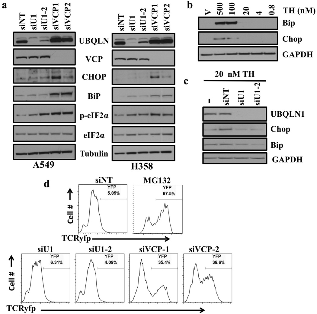
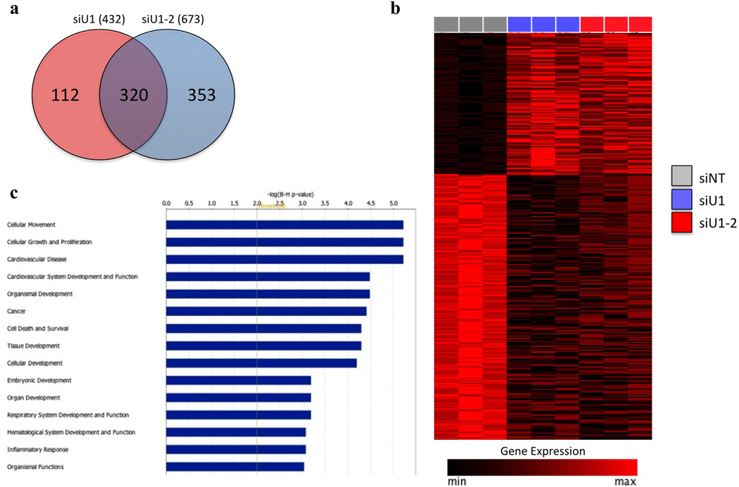
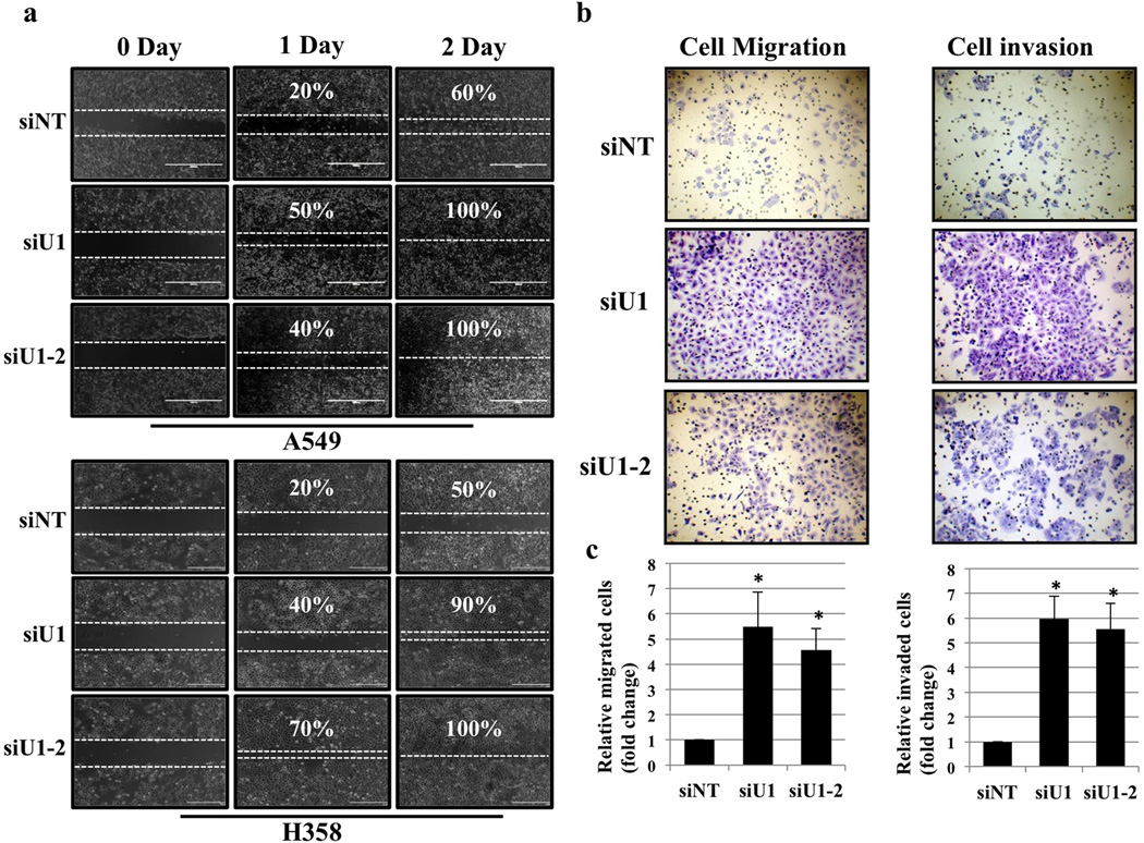
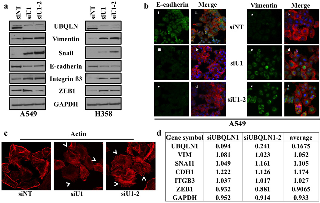
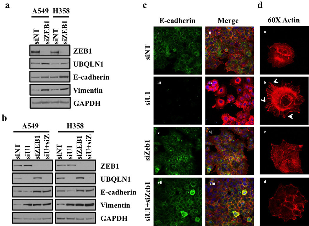
Ubiquilin1 (UBQLN1) is a ubiquitin-like domain and a ubiquitin-associated domain containing protein that has been reported to be involved in shuttling proteins to the proteasome, especially during endoplasmic reticulum-associated protein degradation. Thus, UBQLN1 function has been shown to be critical for combating a number of neurological disorders caused by protein aggregation, such as amyotrophic lateral sclerosis, Alzheimer's disease and Huntington's disease. A role for UBQLN1 in regulating processes involved in tumorigenesis has not been demonstrated. Herein, we show that loss of UBQLN1 causes increased cell migration and invasion, actin cytoskeleton reorganization and induction of epithelial-to-mesenchymal transition (EMT). Loss of UBQLN1 results in a significant decrease in the expression of epithelial markers including E-cadherin and claudin1, whereas expression of mesenchymal markers including Vimentin, Snail and ZEB1 are significantly elevated. Interestingly, we found that ZEB1 is required for induction of mesenchymal-like properties following loss of UBQLN1 and ZEB1 is capable of repressing expression of UBQLN1, suggesting a physiological, reciprocal regulation of EMT by UBQLN1 and ZEB1. Further, we find evidence for a role for UBQLN2 in also regulating EMT and cell migration. These observations have potential clinical relevance because the UBQLN1 gene is lost and underexpressed in a large percentage of human cancer cell lines, and primary human lung cancer samples and recurrent mutations in all five UBQLN family members have been identified in human lung cancers. Taken together, our results suggest for the first time a role for UBQLN family members in cancer biology.


Conflict of interest statement

Conflict of Interest: the authors declare no conflict of interest

Figures

Figure 1
Figure 1. Loss of UBQLN1 does not induce ER stress or alter ERAD
(a) Western blot analysis of proteins involved in unfolded protein response and ER stress response. Cells were transfected with either non targeting (siNT) siRNA or two different siRNAs targeting UBQLN1 (siU1 and siU2) and two different siRNAs targeting VCP (siVCP1 and siVCP2). After 72 hrs of transfection, cells were harvested and analyzed for protein expression. (b) Western Blot analysis of proteins involved in ER stress response. A549 cells were treated with vehicle or with indicated concentrations of Thapsigargin (TH). (c) Western Blot analysis of proteins involved in ER stress response. A549 cells were transfected with either non targeting (siNT) siRNA or two different siRNAs targeting UBQLN1 (siU1 and siU2). 48 hrs post siRNA transfections cells were treated with vehicle or with 20nM of Thapsigargin (TH) for 30 min and harvested 24 hrs later. (d) UBQLN1 loss does not alter ERAD. 293T cells stably expressing TCRyfp, a well-known ERAD substrate, were untreated (NT), treated with MG132, or transfected with the indicated siRNA. Cells were subjected to flow cytometry 24 hrs after MG132 treatment or 72 hrs post siRNA transfection to determine the intensity of yfp signal. MG132 treatment is used as a positive control, as ERAD is known to rely on the proteasome for destruction of substrates.
Figure 2
Figure 2. Inhibition of UBQLN1 in A549 cells induces a gene expression signature related to EMT
(a) Genes differentially expressed upon UBQLN1 knockdown in A549 cells. Comparison of gene expression profiles between A549 cells transfected with either siU1 or siU1-2 and siNT identified a set of 320 unique overlapping genes differentially expressed upon UBQLN1 knockdown (FDR<0.05, fold-change > or < 2). (b) Expression levels of the 320 UBQLN1 regulated genes in siU1, siU2 and siNT treated A549 cells. Genes are arranged in descending order of the average fold change of siU1 and siU1-2 treated cells vs siNT. Expression values are displayed scaled to the maximum expression for each individual gene across the samples. (c) Cellular functions and disease processes enriched within the UBQLN1 gene signature. IPA analysis of the 320 UBQLN1 regulated genes identified cellular movement as the most significantly enriched function. The top 15 diseases and disorders, molecular and cellular functions, and physiological development and function processes are indicated (B-H corrected p-value <0.01).
Figure 3
Figure 3. UBQLN1 loss induces cell migration and invasion
(a) Migration assay in A549 and H358 cells. Cells were transfected with either non targeting (siNT) siRNA or two different siRNAs targeting UBQLN1 (siU1 and siU1-2). 24 hrs post siRNA transfections a pipette tip was used to scratch the dish to make a “wound”. Cells were examined after wound has been formed and successively for 24hr and 48hr post wound formation and photographed. (b) Invasion assay in A549 cells. Cells were transfected with either non targeting (siNT) siRNA or two different siRNAs targeting UBQLN1 (siU1 and siU1-2). 24 hours after siRNA transfection cells were seeded into Boyden chambers without (left) or with (right) matrigel. The lower chamber contained media with serum, whereas the upper chamber containing the cells was without serum. 48 hrs later cells on the underside of the membrane were fixed and stained. (c) Quantification of relative number of cells migrated or invaded through matrigel (*P < 0.05).
Figure 4
Figure 4. Loss of UBQLN1 induces EMT
(a) UBQLN1 loss induces EMT in A549 and H358 cells. A549 and H358 cells were transfected with either with non-targeting siRNA (siNT) or siRNAs targeting UBQLN1 (siU1, siU1-2). After 72 hrs of transfection cells were harvested and analyzed for protein expression using the indicated antibodies. (b) Fluorescence staining for E-cadherin and Vimentin in A549. After 24 hrs of transfection either with non-targeting siRNA (siNT) or with siRNAs targeting UBQLN1 (siU1 and siU1-2) cells were trypsinized and plated on chamber slides and stained for EMT markers. i, iii and v: E-cadherin was detected using Alexa Fluor 488 goat anti-rabbit IgG (green). ii, iv and vi: overlay of respective E-cadherin and F-actin (Alexa Fluor 568 Phalloidin; red) staining with DAPI counter stain. a, c and e: Vimentin was detected using Alexa Fluor 488 goat anti-rabbit IgG (green). b, d and f: overlay of respective E-Vimentin and F-actin (Alexa Fluor 568 Phalloidin; red) staining with DAPI counter stain. (c) Cells were prepared as described in B and F-actin was detected with Alexa Fluor 568 Phalloidin (red). Re-organization of actin cytoskeleton through destruction and cellular protrusion formation is indicated by arrows. (d) Table indicating the fold change of mRNA following siRNA mediated knockdown of UBQLN1 for EMT-associated genes, as compared to non-targeting siRNA transfected cells. Values are in fold change and each value is the average of the triplicate samples for each siRNA.
Figure 5
Figure 5. Coordinate regulation of EMT by UBQLN1 and ZEB1
(a) Loss of Zeb1 increases expression of UBQLN1 and increases epithelial markers in A549 and H358 cells. Western blot analysis of ZEB1, UBQLN1 and EMT markers in A549 and H358 cells. Cells were transfected with either non-targeting siRNA (siNT) or with siRNA targeting ZEB1 (siZEB1). After 72 hrs of transfection, cells were harvested and subjected to western blot for protein expression analysis for UBQLN1, ZEB1 along with other EMT markers (b) UBQLN1 loss requires ZEB1 to induce EMT in A549 and H358 cells. Cells were transfected with non-targeting siRNA, with siUBQLN1, siZEB1 or the combination of siUBQLN1 and siZEB1. After 72 hrs of transfection, cells were harvested. Western blot analysis confirming knockdown of UBQLN1 and ZEB1 along with different EMT markers. (c) Fluorescence staining for E-cadherin in A549. After 24 hrs of transfection either with non-targeting siRNA (siNT) or with siRNAs targeting UBQLN1 (siU1), siZeb1 or combination of siU1 and siZeb1, cells were trypsinized and plated on chamber slides and stained for E-cadherin. i, iii, v and vii: E-cadherin was detected using Alexa Fluor 488 goat anti-rabbit IgG (green). ii, iv, vi and viii: overlay of respective E-cadherin and F-actin (Alexa Fluor 568 Phalloidin; red) staining with DAPI counter stain. (d) A549 cells were prepared as described in (c) and F-actin was detected with Alexa Fluor 568 Phalloidin (red) with 60x objective. Re-organization of actin cytoskeleton through destruction and cellular protrusion formation is indicated by arrows.
Figure 6
Figure 6. UBQLN1 is lost and under-expressed in human cancers
(a) UBQLN1 is frequently lost and underexpressed in cancer cell lines. Cancer cell lines are separated by UBQLN1 copy number status and their corresponding UBQLN1 expression plotted. Box-plots depict the median group expression (middle line), the 25th and 75th percentiles (bottom and top of box, respectively), with the whiskers indicating the 10th and 90th percentiles. Values for all samples outside this range are represented by dots. The UBQLN1 locus is lost in 243 of the 877 cell lines examined. UBQLN1 is significantly under-expressed in samples with UBQLN1 loss or deletion (p<0.0001, Mann-Whitney U-Test). (b) UBQLN1 is frequently lost and underexpressed in human lung adenocarcinomas. Samples are plotted as in A. The UBQLN1 locus is lost in 117 of 230 lung cancers examined. UBQLN1 is significantly underexpressed in tumors with loss or deletion (p<0.0001, Mann-Whitney U-Test). (c) Cancer relevant nonsense mutations in UBQLN1 are degraded in a proteasome dependent manner. Two nonsense mutations found in UBQLN1 in human cancers (UBQLN1G573X and UBQLN1G499X) were introduced into wild-type UBQLN1 (UBQLN1wt). Constructs expressing the indicated gene or an empty vector (V) were transfected into 293t cells and 36 hours post transfection they were treated with MG132 for the indicated times. GFP is expressed from the same mRNA as the UBQLN1 genes following and IRES. (d) Recurrent mutations observed in both UBQLN1 and UBQLN2 which have been identified in human lung cancers. (e) Loss of UBQLN2 induces EMT. UBQLN2 loss induces EMT in A549 and H358 cells. A549 and H358 cells were transfected with either with non-targeting siRNA (siNT) or siRNAs targeting UBQLN2 (siU2-5, siU2-6). After 72 hrs of transfection cells were harvested and analyzed for protein expression using the indicated antibodies. Arrow indicates the UBQLN2 band.
Figure 7
Figure 7. Schematic presentation of regulation of EMT by UBQLN1
See text for details.

References

    1. Lim PJ, Danner R, Liang J, Doong H, Harman C, Srinivasan D, et al. Ubiquilin and p97/VCP bind erasin, forming a complex involved in ERAD. J Cell Biol. 2009;187:201–217. - PMC - PubMed
    1. Lu A, Hiltunen M, Romano DM, Soininen H, Hyman BT, Bertram L, et al. Effects of ubiquilin 1 on the unfolded protein response. J Mol Neurosci. 2009;38:19–30. - PubMed
    1. Kim H, Kim HY, Choi MR, Hwang S, Nam KH, Kim HC, et al. Potentiation of amyotrophic lateral sclerosis (ALS)-associated TDP-43 aggregation by the proteasometargeting factor, ubiquilin 1. J Biol Chem. 2009;284:8083–8092. - PMC - PubMed
    1. Arias-Vásquez A, de Lau L, Pardo L, Liu F, Feng BJ, Bertoli-Avella A, et al. Relationship of the Ubiquilin 1 gene with Alzheimer's and Parkinson's disease and cognitive function. Neurosci Lett. 2007;424:1–5. - PubMed
    1. Olzmann JA, Li L, Chin LS. Aggresome formation and neurodegenerative diseases: therapeutic implications. Curr Med Chem. 2008;15:47–60. - PMC - PubMed

Publication types

MeSH terms