. 2014 Apr 18;9(4):e95229.
doi: 10.1371/journal.pone.0095229. eCollection 2014.

The influence of body position on cerebrospinal fluid pressure gradient and movement in cats with normal and impaired craniospinal communication

Affiliations

The influence of body position on cerebrospinal fluid pressure gradient and movement in cats with normal and impaired craniospinal communication

Marijan Klarica et al. PLoS One. .

Abstract

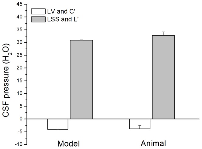
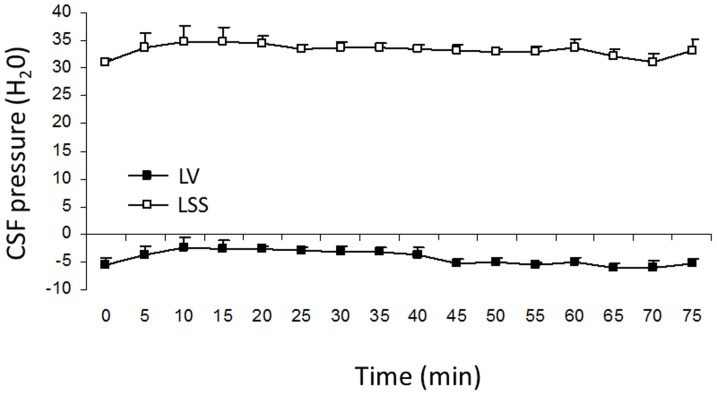
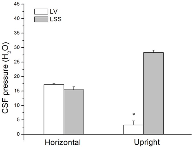
Intracranial hypertension is a severe therapeutic problem, as there is insufficient knowledge about the physiology of cerebrospinal fluid (CSF) pressure. In this paper a new CSF pressure regulation hypothesis is proposed. According to this hypothesis, the CSF pressure depends on the laws of fluid mechanics and on the anatomical characteristics inside the cranial and spinal space, and not, as is today generally believed, on CSF secretion, circulation and absorption. The volume and pressure changes in the newly developed CSF model, which by its anatomical dimensions and basic biophysical features imitates the craniospinal system in cats, are compared to those obtained on cats with and without the blockade of craniospinal communication in different body positions. During verticalization, a long-lasting occurrence of negative CSF pressure inside the cranium in animals with normal cranio-spinal communication was observed. CSF pressure gradients change depending on the body position, but those gradients do not enable unidirectional CSF circulation from the hypothetical site of secretion to the site of absorption in any of them. Thus, our results indicate the existence of new physiological/pathophysiological correlations between intracranial fluids, which opens up the possibility of new therapeutic approaches to intracranial hypertension.


Conflict of interest statement

Competing Interests: The authors have declared that no competing interests exist.

Figures

Figure 1
Figure 1. Scheme of a cat experimental model.
A. 1 – pressure transducer connected to the cannula inside the lateral ventricle; 2 – pressure transducer connected to the cannula inside the lumbar subarachnoid space; 3 – Quand Bridge; 4 – PowerLab/800; 5 – personal computer; hc – distance between the cisterna magna and the pressure measuring point inside the lateral ventricle; hs – distance between the cisterna magna and the pressure measuring point inside the lumbar subarachnoid space. B. Different cat's and model's body positions in which fluid pressure changes have been recorded. “c”- cranial part of the CSF system or cranium in cats; “s” – spinal part of the CSF system or spinal subarachnoid space; horizontal position −0-°; cranium or “cranial” part of the model facing upwards−45-°; cranium or “cranial” part of the model facing upwards −90-° (upright position); cranium or “cranial” part of the model facing downwards−225-°; cranium or “cranial” part of the model facing downwards −270-° (head- down position).
Figure 2
Figure 2. Scheme of a new CSF system model in an upright position.
A. “c” represents a “cranial” part of the model (length 6 cm; gray colour) made from a plastic tube with rigid walls. “s” represents a “spinal” part of the model (length 31 cm) made from a rubber baloon. P1-fluid pressure in the “cranial” part of the model measured via a pressure transducer 1, which is fixed onto the board at an adequate hydrostatic level. Pa is atmospheric pressure, and it represents a reference pressure of 0 cm H2O. P2 is fluid pressure at the distal end of the “spinal” part of the model, recorded via a pressure transducer 2, which is fixed onto the board at the same hydrostatic level. “hc” - distance between open end of the “cranial” part of the model and the pressure measuring point. “hs” – distance between open end of the “cranial” part of the model and the pressure measuring point inside of the “spinal” part of the model. 3 – Quand Bridge; 4 – PowerLab/800; 5 – personal computer. B. Scheme of a “cranial” part of the CSF system model. g – gravitational force; ρ - is fluid density; “hc” - distance between open end of the “cranial” part of the model and the pressure measuring point; P1 – fluid pressure at the top of the plastic tube with open end on its bottom; Pa – atmospheric pressure. C. Scheme of a “spinal” part of the CSF system model. g – gravitational force; ρ - is fluid density; “hs” – distance between open end and the pressure measuring point inside of the “spinal” part of the model; P2 – fluid pressure at the bottom of the “spinal” part of the model; Pa – atmospheric pressure.
Figure 3
Figure 3. Effects of the model position changes (horizontal −0-°; “cranial” part facing upwards −45-°; “cranial” part facing upwards −90-°; “cranial” part facing downwards −225-°; “cranial” part facing downwards −270-°) on the fluid pressure (cm H2O) inside the “cranial” (C') and the “lumbar” (L') part of the model (n = 5).
Results are shown as a mean value ± standard error of the mean (SEM; *p<0.001).
Figure 4
Figure 4. Effects of body position changes (horizontal −0-°, head facing upwards −45-°; head facing upwards−90-°; head facing downwards −225-°; head facing downwards −270-°) on CSF pressures (cm H2O) inside the lateral ventricle (LV), and the lumbar (LSS) subarachnoid spaces in 8 cats.
Results are shown as a mean value ± SEM (*p<0.001).
Figure 5
Figure 5. Fluid pressure inside the “cranial” (C') and the “lumbar” (L') part of the model (n = 5), and CSF pressure inside the lateral ventricle (LV) and lumbar subarachnoid space (LSS) in cats (n = 8) in an upright position.
Results are shown as a mean value ± SEM (p>0.05).
Figure 6
Figure 6. CSF pressure changes inside the lateral ventricle (LV) and the lumbar subarachnoid space (LSS) in cats in upright position.
In five cats (n = 5) CSF pressure was measured during 50 minute period, and in four cats (n = 4) during 75 minute period. Results are shown as a mean value ± SEM.
Figure 7
Figure 7. CSF pressure changes inside the lateral ventricle (LV) and the lumbar subarachnoid space (LSS) in cats with cervical stenosis (n = 5) in horizontal and head facing upwards at −90-° (head-up) position.
Results are shown as a mean value ± SEM (*p<0.001).
Figure 8
Figure 8. Schematic presentation of the hypothesis by which the appearance of negative CSF pressure inside the cranium in an upright position is being explained, without the changes of intracranial fluid volume.
On the right side of the scheme a plastic tube is shown, filled with fluid and open at the lower end (as in the Figure S4b in File S1). Fluid pressure at the top of the tube (P1) is lower than the atmospheric pressure (P2), and it's value corresponds with the hydrostatic fluid column inside the cylinder (formula image
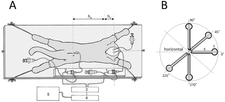
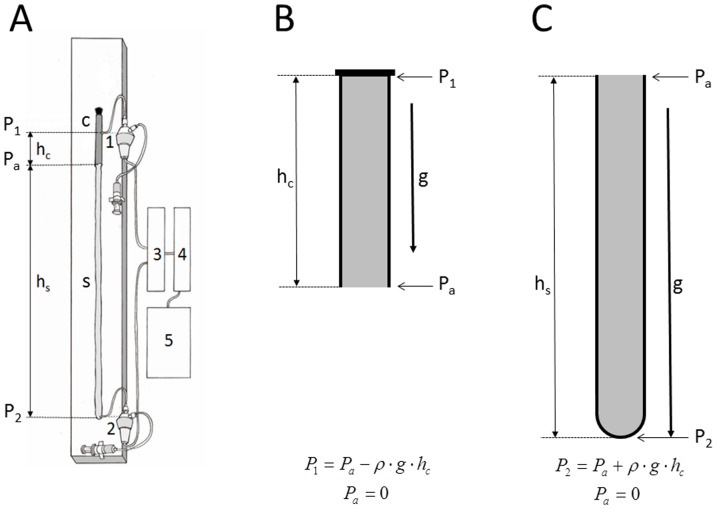
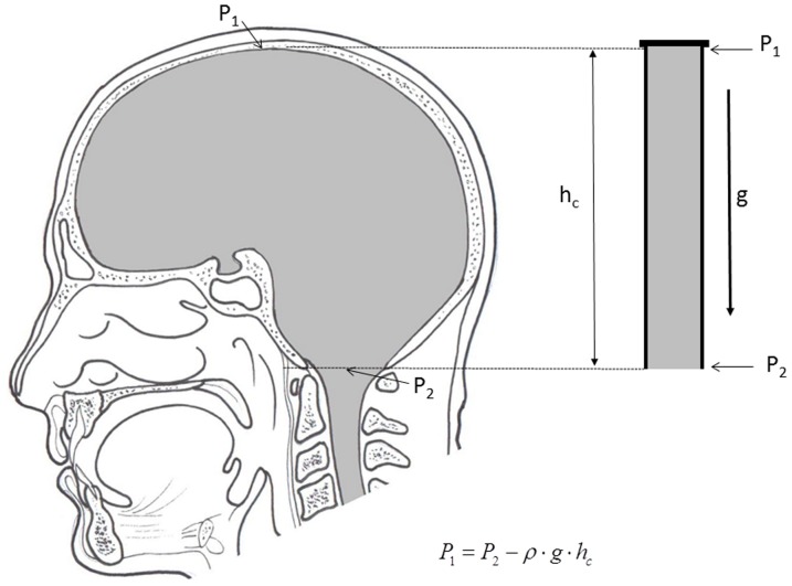
, File S1). Thus, according to the law of fluid mechanics, inside that kind of space negative pressure appears without the changes of the fluid volume. According to the mentioned law, CSF inside the cranium should undergo the same fate (File S1). Namely, according to this law, negative value of the hydrostatic CSF pressure inside the cranium does not depend on the shape of the volume (File S1), but only on the distance between the point of measurement and foramen magnum (hc).

References

    1. Davson H, Welch K, Segal MB (1987) Physiology and pathophysiology of the cerebrospinal fluid. Edinburgh: Churchill-Livingstone. 1013 p.
    1. Pollay M (2010) The function and structure of the cerebrospinal fluid outflow system. Cerebrospinal Fluid Res 21; 7: 9. - PMC - PubMed
    1. Sakka L, Coll G, Chazal J (2011) Anatomy and physiology of cerebrospinal fluid. Europ Ann Otorhinolaryngology, Head Neck Diseases 128: 309–316. - PubMed
    1. Dandy WE (1929) Where is cerebrospinal fluid absorbed? JAMA 92: 2012–2014.
    1. Brierly JF, Field EJ (1948) The connections of the cerebrospinal fluid space with the lymphatic system. J Anat 82: 153–166. - PubMed

Publication types