. 2014 Jun 1;192(11):5415-25.
doi: 10.4049/jimmunol.1400256. Epub 2014 Apr 18.

CD8 T cells use IFN-γ to protect against the lethal effects of a respiratory poxvirus infection

Affiliations

CD8 T cells use IFN-γ to protect against the lethal effects of a respiratory poxvirus infection

John Goulding et al. J Immunol. .

Abstract

CD8 T cells are a key component of immunity to many viral infections. They achieve this through using an array of effector mechanisms, but precisely which component/s are required for protection against a respiratory orthopox virus infection remains unclear. Using a model of respiratory vaccinia virus infection in mice, we could specifically determine the relative contribution of perforin, TRAIL, and IFN-γ-mediated pathways in protection against virus induced morbidity and mortality. Unexpectedly, we observed that protection against death was mediated by IFN-γ without any involvement of the perforin or TRAIL-dependent pathways. IFN-γ mRNA and protein levels in the lung peaked between days 3 and 6 postinfection. This enhanced response coincided with the emergence of virus-specific CD8 T cells in the lung and the cessation of weight loss. Transfer experiments indicated that CD8 T cell-autonomous expression of IFN-γ restricts virus-induced lung pathology and dissemination to visceral tissues and is necessary for clearance of virus. Most significantly, we show that CD8 T cell-derived IFN-γ is sufficient to protect mice in the absence of CD4 and B-lymphocytes. Thus, our findings reveal a previously unappreciated mechanism by which effector CD8 T cells afford protection against a highly virulent respiratory orthopox virus infection.


Figures

Figure 1
Figure 1. Functional IFN-γ signaling is required for limiting viral dissemination and mortality during a respiratory VACV-WR infection
Wild type (WT) C57BL/6J, IFN-γ−/− (a) and IFN-γR−/− (b) mice were intranasally (i.n.) infected with a sub-lethal inoculum of VACV-WR (1.25 × 10⁴ plaque forming units (PFU)/mouse). The animals were weighed daily and euthanized if weight loss was greater than 25 % of their original body weight for two consecutive days. Percentage of initial body weight (a and b; left panels) and mean percent survival (a and b; right panels) from the indicated numbers of mice are presented. Weight loss data are presented as the mean ± SEM of three separate experiments containing 4–8 mice per group and analyzed using a two way ANOVA to determine statistical significance ** p<0.01. Survival data utilizes combined survival data across several experiments using the Mantel-Cox test. Day 7 post infection lung sections stained with hematoxylin and eosin from representative WT and IFN-γ−/− mice (20x magnification) (c). On day 7 post-VACV-WR infection, viral titers (PFU) were determined in the indicated tissues (d). Students t test with Bonferroni’s correction was used to determine viral titer statistical significance, ** p<0.01. WT and IFN-γ−/− mice were also intraperitonealy (i.p.) infected with 2 × 10⁵ PFU VACV-WR and followed for weight loss (e) and tittered for virus (f) on day 8 post infection.
Figure 2
Figure 2. Inflammatory gene expression profile of VACV-WR infected lung tissue
Wild type (WT) C57BL/6J mice were intranasally (i.n.) infected with VACV-WR (1.25 × 10⁴ plaque forming units (PFU)/mouse). Total lung mRNA transcript levels of 26 inflammatory genes were measured at day 0 (naïve), day 3, day 6 and day 9 post infection and presented as a heat map (a) representing the relative value of the gene expression across all samples. G1 – G4 denotes four temporally distinct groups of genes. Fold change in gene expression between day 3 and naïve, day 6 and day 3 and day 9 and day 6 post infection is also represented (b). All array data are presented as the average gene expression level of 4 combined mice per group at each specified time point analyzed. At the indicated time points post infection with VACV-WR, lungs were collected and assayed independently for total IFN-γ mRNA levels (c) and IFN-γ protein levels using ELISA (d). Data are presented as the mean ± one SEM of three independent experiments with 4 mice per group. Students t test with Bonferroni’s correction was used to determine statistical significance compared to day 0 levels ** p<0.01.
Figure 3
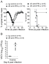
Figure 3. Early IFN-γ signaling is required for survival following a respiratory VACV-WR infection
Wild type (WT) C57BL/6J mice were intranasally (i.n.) infected with VACV-WR (1.25 × 10⁴ plaque forming units (PFU)/mouse). Weight loss (a, left panel) and survival (a, right panel) were monitored in WT mice that were continuously treated with a neutralizing IFN-γ antibody, or isotype control, starting one day before infection (day -1), 3 days or 6 days post i.n. infection with VACV-WR. On day 8 post infection viral titers were measured in mice in which IFN-γ had been neutralized from the outset of infection (b). Data are presented as the mean ± one SEM of two independent experiments with 3–5 mice per group. Survival data utilizes combined survival data across combined experiments using the Mantel-Cox test ** p<0.01.
Figure 4
Figure 4. CD8 T cell priming is quantitatively and qualitatively analogous in wild type and IFN-γ−/− mice
Wild type (WT) C57BL/6J and IFN-γ−/− mice were intranasally (i.n.) infected with 1.25 × 10⁴ plaque forming units (PFU) of VACV-WR. On day 8 post infection, total lung cells were stained with CD3ε, CD8α, CD44, B8R tetramer, intracellular Ki67 or stimulated with B8R peptide and subsequently stained for intracellular IFN-γ and TNF-α. Total numbers (a) of CD3ε+CD8α, CD3ε+CD8α+CD44high, B8R tetramer-positive T cells, relative frequency of CD3ε+CD8α+CD44high B8R tetramer and Ki67 positive cells (b; left and middle panel, respectively) and representative plots for cytokine staining (b; right panel), gated on CD8α+CD62Llow cells, are indicated. Total numbers of day 8 lung CD8α+TNF producing CD8 T cells (c) are also quantified. Quadrant settings were based on controls, using infected cells that were not stimulated with peptide or stained with B8R tetramer, and uninfected lung cells stimulated with B8R peptide (data not shown). Results are mean number ± SEM (n = 4 mice/group) from one experiment. Similar results were obtained in three separate experiments. In situ CD8 T cell location (peribronchial and parenchymal) was also determined in naïve and day 8 infected OCT embedded lung sections fluorescently stained for actin (green) and CD8α (magenta) (d – f).
Figure 5
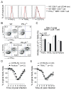
Figure 5. CD8 T cell expressed cytolytic effector molecules provide limited protection during a respiratory VACV-WR infection
Wild type (WT) C57BL/6J and IFN-γ−/− mice were intranasally (i.n.) infected with 1.25 × 10⁴ plaque forming units (PFU) of VACV-WR. On day 7 post infection, total lung B8R positive CD8 T cells were stained for surface expression of FASL and TRAIL (a). Naïve CD8+CD44low B8R-tetramer negative cells were used as controls. The surface expression of CD107α (a; right panel) was also determined in day 7 post infection WT and IFN-γ−/− following overnight B8R peptide stimulation of total lung cells and in combination with intracellular cytokine (IFN-γ) staining (b). In addition, day 7 B8R-tetramer positive lung CD8 T cells were FACS sorted and examined for mRNA levels for FASL, TRAIL, granzyme B (Grz-B) and perforin which were normalized to the expression of L32 and GAPDH housekeeper genes (c). Results are mean number ± SEM (n = 4 mice/group) from one experiment. Similar results were obtained in two separate experiments. Perforin-deficient (perforin−/−; C57BL/6J) (d), TRAIL-deficient TRAIL−/−; C57BL/6J) (e) and WT C57BL/6J mice were intranasally (i.n.) infected with VACV-WR (1.25 × 10⁴ PFU/mouse). The animals were weighed daily and euthanized if weight loss was greater than 25 % of their original body weight for two consecutive days. Percentage of initial body weight (d and e) from the indicated numbers of mice are presented. Data are presented as the mean ± one SEM of three independent experiments with 4–8 mice per group.
Figure 6
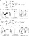
Figure 6. IFN-γ competent CD8 T cells can protect lymphopenic but not IFN-γR−/− mice against mortality following a respiratory VACV-WR infection
A total of 5 × 10⁶ naive (CD3+CD8+CD44low) WT or IFN-γ −/− CD8 T cells were transferred i.v. into RAG−/− mice that were infected intranasally (i.n.) 24 h later with 1.25 × 10⁴ plaque forming units (PFU) VACV-WR (a). WT and RAG−/− mice with no CD8 T cells transferred were used as controls. The animals were weighed daily and euthanized if weight loss was greater than 25 % of their original body weight for two consecutive days. Percentage of initial body weight (b; left panel) and mean percent survival (b; right panel) from the indicated numbers of mice are presented. On day 21 post infection, total lung cells from WT and RAG−/− mice that received WT CD8 T cells were stimulated with B8R peptide and assessed for intracellular IFN-γ production (c). To determined frequency of cytokine producing cells CD8 T cells were gated on CD8+CD62Llow cells and a representative FACS plot from each surviving experimental group was shown. In a separate experiment, 5 × 10⁶ naive (CD3+CD8+CD44low) WT CD8 T cells were transferred i.v. into IFN-γR−/− mice that were infected i.n. 24 h later with VACV-WR (d). Percentage of initial body weight (e; left panel) and mean percent survival (e; right panel) from the indicated numbers of mice are presented. On day 8 post infection total lung cells were isolated and stimulated with B8R peptide to determine the frequency of cytokine producing CD8 T cells (f). Weight loss data are presented as the mean ± SEM of three separate experiments containing 3–5 mice per group and analyzed using a two way ANOVA to determine statistical significance ** p<0.01. Survival data utilizes combined survival data across combined experiments using the Mantel-Cox test.
Figure 7
Figure 7. CD8 T cell derived IFN-γ is sufficient for protection in the absence of host derived IFN-γ
A total of 5 × 10⁶ naive (CD3+CD8+CD44low) WT CD8 T cells were transferred i.v. into IFN-γ−/− mice that were infected i.n. 24 h later with VACV-WR (a). The animals were weighed daily and euthanized if weight loss was greater than 25 % of their original body weight for two consecutive days. Percentage of initial body weight (b; left panel) and mean percent survival (b; right panel) from the indicated numbers of mice are presented. WT and IFN-γ−/− mice with no T cell transfer were used as control. On day 21 post infection, lungs were harvested and stimulated overnight with B8R peptide for intracellular IFN-γ and TNF staining (c). Representative plots for cytokine staining, gating on CD8+CD62Llow cells, are shown and the percentages that stained positive for IFN-γ alone or TNF and IFN-γ/TNF are indicated as well as the transferred cells (IFN-γ+) in IFN-γ−/− mice. Data are presented as the mean ± SEM of three separate experiments containing 3–5 mice per group; weight loss data was analyzed using a two way ANOVA to determine statistical significance. Survival data utilizes combined survival data across combined experiments using the Mantel-Cox test ** p<0.01.

Similar articles

Cited by

References

    1. Moss B. Poxviridae: the viruses and their replication. In: Knipe DM, editor. Fields Virology. 5. Lippincott Williams & Wilkins; Philadelphia: 2007. pp. 2905–2946.
    1. Cann JA, Jahrling PB, Hensley LE, Wahl-Jensen V. Comparative pathology of smallpox and monkeypox in man and macaques. J Comp Pathol. 2013;148:6–21. - PMC - PubMed
    1. Arita I, Jezek Z, Khodakevich L, Ruti K. Human monkeypox: a newly emerged orthopoxvirus zoonosis in the tropical rain forests of Africa. The American journal of tropical medicine and hygiene. 1985;34:781–789. - PubMed
    1. Di Giulio DB, Eckburg PB. Human monkeypox: an emerging zoonosis. Lancet Infect Dis. 2004;4:15–25. - PMC - PubMed
    1. Baxby D, Bennett M, Getty B. Human cowpox 1969–93: a review based on 54 cases. The British journal of dermatology. 1994;131:598–607. - PubMed

Publication types

MeSH terms

Substances