Developmental change in the function of movement systems: transition of the pectoral fins between respiratory and locomotor roles in zebrafish
- PMID: 24748600
- PMCID: PMC4097112
- DOI: 10.1093/icb/icu014
Developmental change in the function of movement systems: transition of the pectoral fins between respiratory and locomotor roles in zebrafish
Abstract
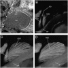
An animal may experience strikingly different functional demands on its body's systems through development. One way of meeting those demands is with temporary, stage-specific adaptations. This strategy requires the animal to develop appropriate morphological states or physiological pathways that address transient functional demands as well as processes that transition morphology, physiology, and function to that of the mature form. Recent research on ray-finned (actinopterygian) fishes is a developmental transition in function of the pectoral fin, thereby providing an opportunity to examine how an organism copes with changes in the roles of its morphology between stages of its life history. As larvae, zebrafish alternate their pectoral fins in coordination with the body axis during slow swimming. The movements of their fins do not appear to contribute to the production of thrust or to stability but instead exchange fluid near the body for cutaneous respiration. The morphology of the larval fin includes a simple stage-specific endoskeletal disc overlaid by fan-shaped adductor and abductor muscles. In contrast, the musculoskeletal system of the mature fin consists of a suite of muscles and bones. Fins are extended laterally during slow swimming of the adult, without the distinct, high-amplitude left-right fin alternation of the larval fin. The morphological and functional transition of the pectoral fin occurs through juvenile development. Early in this period, at about 3 weeks post-fertilization, the gills take over respiratory function, presumably freeing the fins for other roles. Kinematic data suggest that the loss of respiratory function does not lead to a rapid switch in patterns of fin movement but rather that both morphology and movement transition gradually through the juvenile stage of development. Studies relating structure to function often focus on stable systems that are arguably well adapted for the roles they play. Examining how animals navigate transitional periods, when the link of structure to function may be less taut, provides insight both into how animals contend with such change and into the developmental pressures that shape mature form and function.
© The Author 2014. Published by Oxford University Press on behalf of the Society for Integrative and Comparative Biology.
Figures


Similar articles
-
Movement and function of the pectoral fins of the larval zebrafish (Danio rerio) during slow swimming.J Exp Biol. 2011 Sep 15;214(Pt 18):3111-23. doi: 10.1242/jeb.057497. J Exp Biol. 2011. PMID: 21865524
-
Swimming of larval zebrafish: fin-axis coordination and implications for function and neural control.J Exp Biol. 2004 Nov;207(Pt 24):4175-83. doi: 10.1242/jeb.01285. J Exp Biol. 2004. PMID: 15531638
-
Development of zebrafish (Danio rerio) pectoral fin musculature.J Morphol. 2005 Nov;266(2):241-55. doi: 10.1002/jmor.10374. J Morphol. 2005. PMID: 16163704
-
The Water to Land Transition Submerged: Multifunctional Design of Pectoral Fins for Use in Swimming and in Association with Underwater Substrate.Integr Comp Biol. 2022 Oct 29;62(4):908-921. doi: 10.1093/icb/icac061. Integr Comp Biol. 2022. PMID: 35652788 Free PMC article. Review.
-
Fins as Mechanosensors for Movement and Touch-Related Behaviors.Integr Comp Biol. 2018 Nov 1;58(5):844-859. doi: 10.1093/icb/icy065. Integr Comp Biol. 2018. PMID: 29917043 Review.
Cited by
-
Acquired versus innate prey capturing skills in super-precocial live-bearing fish.Proc Biol Sci. 2016 Jul 13;283(1834):20160972. doi: 10.1098/rspb.2016.0972. Proc Biol Sci. 2016. PMID: 27412277 Free PMC article.
-
Transformation of an early-established motor circuit during maturation in zebrafish.Cell Rep. 2022 Apr 12;39(2):110654. doi: 10.1016/j.celrep.2022.110654. Cell Rep. 2022. PMID: 35417694 Free PMC article.
-
Temporal Vestibular Deficits in synaptojanin 1 (synj1) Mutants.Front Mol Neurosci. 2021 Jan 18;13:604189. doi: 10.3389/fnmol.2020.604189. eCollection 2020. Front Mol Neurosci. 2021. PMID: 33584199 Free PMC article.
-
A primal role for the vestibular sense in the development of coordinated locomotion.Elife. 2019 Oct 8;8:e45839. doi: 10.7554/eLife.45839. Elife. 2019. PMID: 31591962 Free PMC article.
-
Differential expression and hypoxia-mediated regulation of the N-myc downstream regulated gene family.FASEB J. 2021 Nov;35(11):e21961. doi: 10.1096/fj.202100443R. FASEB J. 2021. PMID: 34665878 Free PMC article.
References
-
- Asaduzzaman MD, Akolkar DB, Kinoshita S, Watabe S. The expression of multiple myosin heavy chain genes during skeletal muscle development of torafugu Takifugu rubripes embryos and larvae. Gene. 2013;515:144–54. - PubMed
-
- Bardach J, Case J. Sensory capabilities of the modified fins of squirrel hake (Urophycis chuss) and searobins (Prionotus carolinus and P. evolans) Copeia. 1965;1965:194–206.
-
- Barrionuevo WR, Burggren WW. O2 consumption and heart rate in developing zebrafish (Danio rerio): influence of temperature and ambient O2. Am J Physiol. 1999;276:R505–13. - PubMed
-
- Blake RW. Influence of pectoral fin shape on thrust and drag in labriform locomotion. J Zool Lond. 1981;194:53–66.
Publication types
MeSH terms
LinkOut - more resources
Full Text Sources
Other Literature Sources
Research Materials

