. 2014 Jul;196(13):2376-86.
doi: 10.1128/JB.01563-14. Epub 2014 Apr 18.

Txc, a new type II secretion system of Pseudomonas aeruginosa strain PA7, is regulated by the TtsS/TtsR two-component system and directs specific secretion of the CbpE chitin-binding protein

Affiliations

Txc, a new type II secretion system of Pseudomonas aeruginosa strain PA7, is regulated by the TtsS/TtsR two-component system and directs specific secretion of the CbpE chitin-binding protein

Frédéric Cadoret et al. J Bacteriol. 2014 Jul.

Abstract

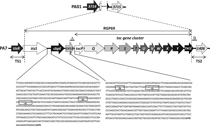
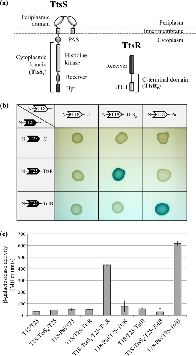
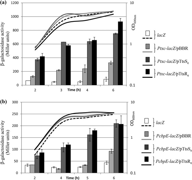
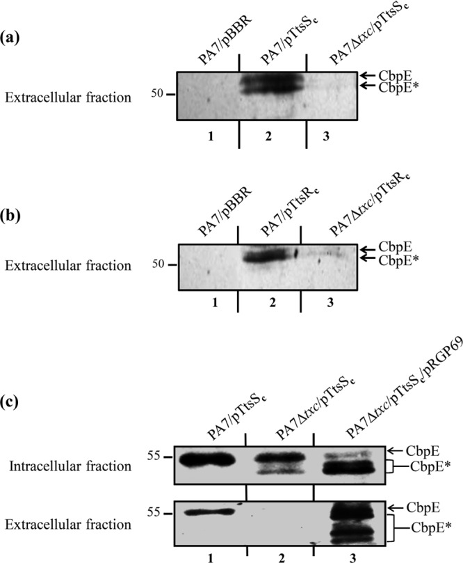
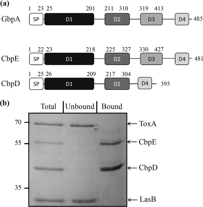
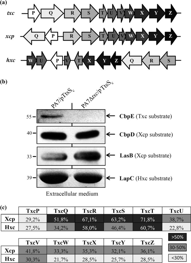
We present here the functional characterization of a third complete type II secretion system (T2SS) found in newly sequenced Pseudomonas aeruginosa strain PA7. We call this system Txc (third Xcp homolog). This system is encoded by the RGP69 region of genome plasticity found uniquely in strain PA7. In addition to the 11 txc genes, RGP69 contains two additional genes encoding a possible T2SS substrate and a predicted unorthodox sensor protein, TtsS (type II secretion sensor). We also identified a gene encoding a two-component response regulator called TtsR (type II secretion regulator), which is located upstream of the ttsS gene and just outside RGP69. We show that TtsS and TtsR constitute a new and functional two-component system that controls the production and secretion of the RGP69-encoded T2SS substrate in a Txc-dependent manner. Finally, we demonstrate that this Txc-secreted substrate binds chitin, and we therefore name it CbpE (chitin-binding protein E).


Figures

FIG 1
FIG 1
Genetic organization of PA7 RGP69. The genomic island RGP69, uniquely present in P. aeruginosa strain PA7, possesses 14 predicted open reading frames encoding the 11 putative T2SS Txc components (TxcP to TxcZ) and 3 genes, a gene encoding a predicted chitin-binding protein (CbpE), a gene encoding an unorthodox sensor protein (TtsS), and PSPA7_1418, which is predicted to encode a protein belonging to the cytochrome b superfamily. RGP69 is externally flanked by the ttsR and PSPA7_1406 (1406) genes, which are homologous to the two contiguous genes PA3714 (3714) and PA3715 (3715), respectively, in strain PAO1. The two DNA sequences corresponding to the segments used to construct the PcbpE-lacZ and PtxcP-lacZ reporter fusions are also shown. The −10 and −35 boxes of the two predicted sigma 70 promoter regions of RGP69 are boxed in the corresponding DNA sequences. The ATG start codons of cbpE and txcP are indicated in boldface type. Also indicated are the two DNA regions TS1 and TS2, used to clone the RGP69 sequence in the gene capture vector pLLX13. A and B regions correspond to the DNA sequences (500 bp) cloned into pKNG208 to generate the suicide vector pKNG208Δtxc, which was used to generate the txc deletion mutant.
FIG 2
FIG 2
TtsS and TtsR interact together to form a two-component system. (a) Modular structures of the unorthodox sensor TtsS and the response regulator TtsR. The cytoplasmic domain of TtsS (TtsSc) and the C-terminal domain of TtsR (TtsRc) used for transcriptional and secretion experiments are indicated by brackets. All the other characteristic domains of the unorthodox sensor and NarL-type response regulator are indicated (Hpt for histidine phosphotransferase and HTH for helix-turn-helix). (b) Binary interactions between TtsSc and full-length TtsR determined by using the BACTH assay. TtsSc and TtsR were fused to the C termini of Bordetella pertussis adenylate cyclase fragments T18 and T25, leading to plasmids pT18-TtsSc and pT25-TtsR. Various T18 and T25 plasmid combinations were cotransformed into E. coli cya strain BTH101 and plated onto LB agar–IPTG–X-gal medium. Functional complementation between the T18 and T25 fragments, which occurs only upon interactions of the hybrid proteins, triggers the expression of the cya gene and yields blue colonies. The E. coli TolB and Pal interacting proteins were used as a positive control. (c) Interactions were quantified by β-galactosidase assays in biological triplicates for all tested interactions.
FIG 3
FIG 3
The TtsS/TtsR two-component system positively regulates cbpE and txc gene expressions. Shown are β-galactosidase activities of Ptxc-lacZ (a) and PcbpE-lacZ (b) transcriptional fusions (in Miller units) in PA7::Ptxc-lacZ (Ptxc-lacZ) and PA7::PcbpE-lacZ (PcbpE-lacZ) strains overproducing TtsSc or TtsRc or not (pBBR). β-Galactosidase activities were also measured in the strain bearing the promoterless lacZ fusion, PA7::lacZ (lacZ), as a negative control. The growth curves of the strains are also shown (OD600). The experiment was reproduced three times. Samples were duplicated for each experiment, and the corresponding standard deviations were calculated.
FIG 4
FIG 4
Txc-dependent secretion of CbpE by strain PA7 overproducing TtsSc or TtsRc. (a and b) Immunoblots of extracellular protein fractions from wild-type PA7 (PA7) or Txc-deficient PA7 (PA7 Δtxc) carrying the empty vector (pBBR), pTtsSc (a), or pTtsRc (b), probed with anti-CbpE antibody. (c) Immunoblots of intracellular (top) and extracellular (bottom) fractions from wild-type PA7 (PA7) and the Txc-deficient PA7 strain (PA7 Δtxc) carrying pTtsSc alone or pTtsSc and pRGP69, probed with anti-CbpE antibody. CbpE and degradation products of CbpE are indicated by asterisks. Molecular mass markers (in kDa) are indicated on the left.
FIG 5
FIG 5
CbpE is a chitin-binding protein. (a) Schematic representation of the presence of the functional domains of GbpA (GI:15601566) in CbpE (PSPA7_1419) and CbpD (PSPA7_4667). The domains represented are the signal peptide (SP), the GbpA D1 chitin- and mucin-binding domain, the GbpA D2 and D3 bacterial surface-binding domains, and the GbpA D4 chitin-binding domain. Signal peptide domains were predicted by the SignalP 4.1 server (http://www.cbs.dtu.dk/services/SignalP/). When present, the D1, D2, and D3 domains were predicted by homology modeling of CbpE and CbpD based on the structure of GbpA (PDB accession number 2XWX), using Modeler software (68). The D4 domain in CbpE and CbpD was defined based on a sequence alignment of the C-terminal sequences of CbpE, CbpD, and GbpA by using the Multalin Web server (http://multalin.toulouse.inra.fr/multalin/multalin.html). (b) To test the chitin-binding affinity of CbpE, a mixture of affinity-purified CbpE and CbpD and purified LasB and ToxA exoproteins (Total) was incubated with chitin beads according to the protocol described in Materials and Methods. Incubation with chitin beads resulted in the precipitation of the exoproteins interacting with chitin (Bound), while the exoproteins without affinity for chitin remained in the soluble fraction (Unbound). Samples were analyzed by SDS-PAGE followed by Coomassie blue staining. Molecular mass markers (in kDa) are indicated on the left.
FIG 6
FIG 6
Coexistence of three functional T2SSs in strain PA7. (a) Genetic organization of the three T2SS gene clusters in PA7. (b) Comparative analysis of PA7 T2SS secretomes. The extracellular protein fractions of PA7 strains were analyzed by SDS-PAGE followed by Coomassie blue staining for LapC secretion by the Hxc T2SS (LapC identity was confirmed by mass spectrometry), immunodetection with anti-CbpE for CbpE secretion by the Txc T2SS, and immunodetection with anti-CbpD and anti-LasB for CbpD and LasB secretion by the Xcp T2SS. Molecular mass markers (in kDa) are indicated on the left. (c) Sequence relatedness between Txc components and their counterparts in the Xcp and Hxc T2SSs. The percent identities over the complete protein sequences are indicated. Low (<30%), medium (30 to 50%), and high (>50%) identities are shown.

Similar articles

Cited by

References

    1. Driscoll JA, Brody SL, Kollef MH. 2007. The epidemiology, pathogenesis and treatment of Pseudomonas aeruginosa infections. Drugs 67:351–368. 10.2165/00003495-200767030-00003 - DOI - PubMed
    1. Kipnis E, Sawa T, Wiener-Kronish J. 2006. Targeting mechanisms of Pseudomonas aeruginosa pathogenesis. Med. Mal. Infect. 36:78–91. 10.1016/j.medmal.2005.10.007 - DOI - PubMed
    1. Jimenez PN, Koch G, Thompson JA, Xavier KB, Cool RH, Quax WJ. 2012. The multiple signaling systems regulating virulence in Pseudomonas aeruginosa. Microbiol. Mol. Biol. Rev. 76:46–65. 10.1128/MMBR.05007-11 - DOI - PMC - PubMed
    1. Rodrigue A, Quentin Y, Lazdunski A, Mejean V, Foglino M. 2000. Two-component systems in Pseudomonas aeruginosa: why so many? Trends Microbiol. 8:498–504. 10.1016/S0966-842X(00)01833-3 - DOI - PubMed
    1. Bleves S, Viarre V, Salacha R, Michel GP, Filloux A, Voulhoux R. 2010. Protein secretion systems in Pseudomonas aeruginosa: a wealth of pathogenic weapons. Int. J. Med. Microbiol. 300:534–543. 10.1016/j.ijmm.2010.08.005 - DOI - PubMed

MeSH terms

Associated data