. 2014 Jul;196(13):2367-75.
doi: 10.1128/JB.01552-14. Epub 2014 Apr 18.

Sulfolobus replication factor C stimulates the activity of DNA polymerase B1

Affiliations

Sulfolobus replication factor C stimulates the activity of DNA polymerase B1

Xuanxuan Xing et al. J Bacteriol. 2014 Jul.

Abstract

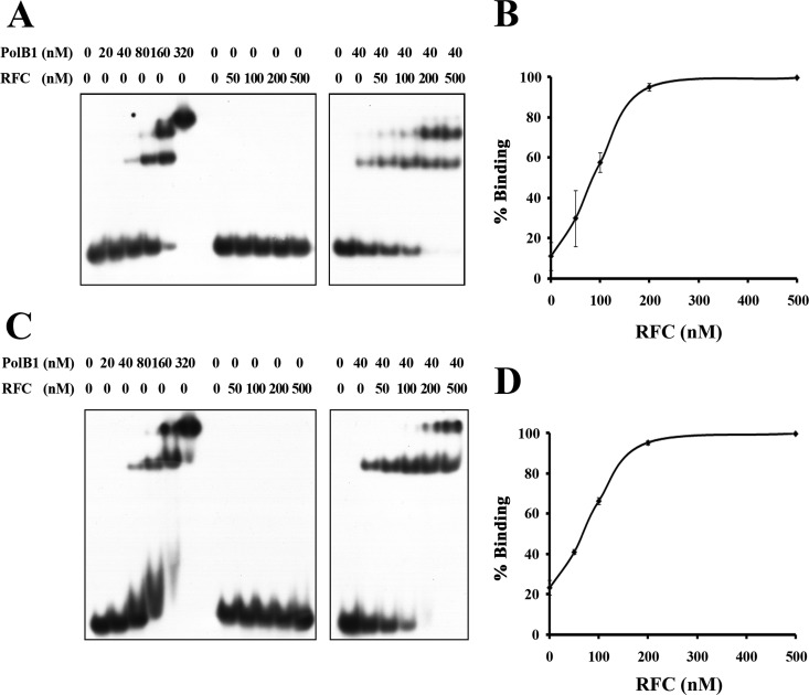
Replication factor C (RFC) is known to function in loading proliferating cell nuclear antigen (PCNA) onto primed DNA, allowing PCNA to tether DNA polymerase for highly processive DNA synthesis in eukaryotic and archaeal replication. In this report, we show that an RFC complex from the hyperthermophilic archaea of the genus Sulfolobus physically interacts with DNA polymerase B1 (PolB1) and enhances both the polymerase and 3'-5' exonuclease activities of PolB1 in an ATP-independent manner. Stimulation of the PolB1 activity by RFC is independent of the ability of RFC to bind DNA but is consistent with the ability of RFC to facilitate DNA binding by PolB1 through protein-protein interaction. These results suggest that Sulfolobus RFC may play a role in recruiting DNA polymerase for efficient primer extension, in addition to clamp loading, during DNA replication.


Figures

FIG 1
FIG 1
Interaction between RFC and PolB1. (A) Pulldown analysis of the interaction between RFC and PolB1 from S. islandicus. S. islandicus (PolB1-Tag) was grown to the exponential phase and harvested. The cell extract was incubated with Ni2+ beads. The beads were subsequently washed and treated with DNase I, if indicated. Proteins retained by the beads were subjected to SDS-PAGE and identified by immunoblotting with antibodies against RFCS. Lane C, a cell extract from S. islandicus, instead of S. islandicus PolB1-Tag, was used in the assay as a control for nonspecific binding of RFC to Ni2+ beads. (B) Blue native PAGE. RFC (30 pmol) and PolB1 (30 pmol) were mixed individually or in combination with loading buffer for blue native PAGE. Samples were subjected to electrophoresis in a 5 to 15% gradient blue native polyacrylamide gel at 10°C. Molecular mass markers are indicated on the left. RFC-PolB1 complexes are indicated by arrows. (C) Bio-layer interferometry. Amine-reactive (AR) biosensors, covered with immobilized PolB1, were treated in RFC solutions of increasing concentration (160, 320, 650, 1,300, and 2,600 nM), as represented by curves from bottom to top, respectively. The rate constants ka and kd were determined, and the equilibrium dissociation constant KD was calculated as described by the manufacturer.
FIG 2
FIG 2
RFC stimulates the polymerase activity of PolB1 in an ATP-independent manner. (A) Effect of RFC on primer extension by PolB1 on an oligonucleotide template. PolB1 (4 nM) was incubated for 15 min at 65°C with 32P-labeled p42/t76 (2 nM) in the presence of indicated amounts of RFC. Samples were extracted with phenol-chloroform and subjected to electrophoresis in a 15% polyacrylamide gel containing 8 M urea in 1× TBE. The gel was exposed to X-ray film. (B) Effect of RFC on primer extension by PolB1 on an M13 ssDNA template. PolB1 (20 nM) was incubated for 15 min at 65°C with 32P-labeled p42/M13 ssDNA (2 nM) in the presence or absence of RFC. Samples were extracted with phenol-chloroform and subjected to electrophoresis in a 1.2% alkaline agarose gel in 50 mM NaOH and 1 mM EDTA. The gel was dried and exposed to X-ray film. One-kilobase size markers with the indicated sizes are on the left. (C) Effect of ATP or ATP-γ-S on stimulation of PolB1 activity by RFC. PolB1 (4 nM) and the indicated amounts of RFC was incubated for 15 min at 65°C with 32P-labeled p42/t76 (2 nM) in the presence or absence of ATP or ATP-γ-S (1 mM). Samples were processed as described above. Left and right panels, reactions were performed at the same time and run on two separate gels under identical conditions. (D and E) Quantitative analysis. Samples, prepared as described for panels A and C, respectively, were subjected to electrophoresis in 15% polyacrylamide gels containing 8 M urea in 1× TBE. The gels were analyzed using a phosphorimager. The data shown represent averages of three independent measurements.
FIG 3
FIG 3
RFC stimulates the exonuclease activity of PolB1. (A) Effect of RFC on DNA cleavage by PolB1. PolB1 (4 nM) was incubated with 32P-labeled p42, p42/t42, or p42/t76 (2 nM) in the presence of the indicated amounts of RFC for 15 min at 65°C. Samples were extracted with phenol-chloroform and subjected to electrophoresis in an 8% polyacrylamide gel containing 8 M urea in 1× TBE. The gel was exposed to X-ray film. Left and right panels, reactions were performed at the same time and run on two separate gels under identical conditions. p42, p42/t42, and p42/t76 are shown as ssDNA, dsDNA, and p/tDNA, respectively. (B) Quantitative analysis. Samples, prepared as described above, were subjected to electrophoresis in an 8% polyacrylamide gel containing 8 M urea in 1× TBE. DNA cleavage was quantified using a phosphorimager. The data shown represent averages of three independent measurements.
FIG 4
FIG 4
Effect of RFCS on the activities of PolB1. (A) Effect of RFCS on primer extension by PolB1. PolB1 (2 nM) was incubated for 15 min at 65°C with 32P-labeled p42/t76 (2 nM) in the presence or absence of RFCS or RFCSL (75 nM). Samples were extracted with phenol-chloroform, and subjected to electrophoresis in a 15% polyacrylamide gel containing 8 M urea in 1× TBE. The gel was exposed to X-ray film. Lanes from the same gel are juxtaposed, as indicated by a thin black line. (B) Effect of RFCS on DNA cleavage by PolB1. PolB1 (2 nM) was incubated for 15 min at 65°C with 32P-labeled p42/t76 (2 nM) in the presence or absence of RFCs or RFCSL (75 nM). Samples were processed as described above.
FIG 5
FIG 5
Stimulation of the PolB1 activity by RFC is independent of the ability of RFC to bind DNA. (A and B) Bio-layer interferometry. Streptavidin biosensors, covered with immobilized 5′-biotinylated t76 annealed to p42, were treated with wild-type (WT) or mutant (MUT) RFC solutions of increasing concentration (20, 39, 78, 156, and 312 nM), as represented by curves from bottom to top, respectively. The rate constants ka and kd were determined, and the equilibrium dissociation constant KD was calculated as described by the manufacturer. (C) Binding of singly nicked pUC18 by RFC. Wild-type or mutant RFC (5 pmol) was mixed with singly nicked pUC18 (5 pmol) in the presence of ATP-γ-S (4 mM). Following incubation for 10 min at 70°C, the mixture was applied to a modified G50 column. Fractions in the void volume were collected, and an aliquot of each fraction was subjected to SDS-PAGE, followed by immunoblotting with anti-RFCS antibodies. Lane C, positive control. (D) Comparison of the effects of wild-type and mutant RFC on primer extension by PolB1. PolB1 (4 nM) was incubated for 15 min at 65°C with 32P-labeled p42/t76 (4 nM) in the presence of the indicated amounts of wild-type or mutant RFC. Samples were extracted with phenol-chloroform and subjected to electrophoresis in an 8% polyacrylamide gel containing 8 M urea in 1× TBE. The gel was exposed to X-ray film. (E) Quantitative analysis. Samples, prepared as described for panel D, were subjected to electrophoresis in an 8% polyacrylamide gel containing 8 M urea in 1× TBE. The gel was analyzed using a phosphorimager. The data shown represent averages of three independent measurements. (F) Comparison of the effects of wild-type and mutant RFC on the exonuclease activity of PolB1. PolB1 (4 nM) was incubated for 15 min at 65°C with 32P-labeled p42/t76 (4 nM) in the presence of the indicated amounts of wild-type or mutant RFC. Samples were processed as described for panel D. (G) Quantitative analysis. Samples, prepared as described for panel F, were subjected to electrophoresis in an 8% polyacrylamide gel containing 8 M urea in 1× TBE. The gel was analyzed using a phosphorimager. The data shown represent averages of three independent measurements.
FIG 6
FIG 6
RFC promotes DNA binding by PolB1. (A) Effect of RFC on the binding of PolB1 to p/tDNA. PolB1 was mixed with 32P-labeled p42/t76 (2 nM) in the presence or absence of RFC. The mixtures were subjected to electrophoresis in an 8% polyacrylamide gel in 0.1× TBE. The gel was exposed to X-ray film. Left and right panels, samples were assembled at the same time and run on two separate gels under identical conditions. (B) Quantitative analysis. Samples were processed as described above. The gel was analyzed using a phosphorimager. The data shown represent averages of three independent measurements. (C) Effect of RFC on the binding of PolB1 to ssDNA. The assays were carried out as described for panel A except that 32P-labeled p42, instead of 32P-labeled p42/t76, was used. Left and right panels, samples were assembled at the same time and run on two separate gels under the identical conditions. (D) Quantitative analysis. Samples were processed as described for panel C. The gel was analyzed using a phosphorimager. The data shown represent averages of three independent measurements.

Similar articles

References

    1. Pomerantz RT, O'Donnell M. 2007. Replisome mechanics: insights into a twin DNA polymerase machine. Trends Microbiol. 15:156–164. 10.1016/j.tim.2007.02.007 - DOI - PubMed
    1. O'Donnell M, Kuriyan J. 2006. Clamp loaders and replication initiation. Curr. Opin. Struct. Biol. 16:35–41. 10.1016/j.sbi.2005.12.004 - DOI - PubMed
    1. Jeruzalmi D, O'Donnell M, Kuriyan J. 2001. Crystal structure of the processivity clamp loader gamma (γ) complex of E. coli DNA polymerase III. Cell 106:429–441. 10.1016/S0092-8674(01)00463-9 - DOI - PubMed
    1. Henneke G, Gueguen Y, Flament D, Azam P, Querellou J, Dietrich J, Hubscher U, Raffin JP. 2002. Replication factor C from the hyperthermophilic archaeon Pyrococcus abyssi does not need ATP hydrolysis for clamp-loading and contains a functionally conserved RFC PCNA-binding domain. J. Mol. Biol. 323:795–810. 10.1016/S0022-2836(02)01028-8 - DOI - PubMed
    1. Matsumiya S, Ishino S, Ishino Y, Morikawa K. 2002. Physical interaction between proliferating cell nuclear antigen and replication factor C from Pyrococcus furiosus. Genes Cells 7:911–922. 10.1046/j.1365-2443.2002.00572.x - DOI - PubMed

Publication types

MeSH terms