. 2014 Jun;42(10):6232-42.
doi: 10.1093/nar/gku274. Epub 2014 Apr 19.

The forkhead transcription factor FOXK2 acts as a chromatin targeting factor for the BAP1-containing histone deubiquitinase complex

Affiliations

The forkhead transcription factor FOXK2 acts as a chromatin targeting factor for the BAP1-containing histone deubiquitinase complex

Zongling Ji et al. Nucleic Acids Res. 2014 Jun.

Abstract

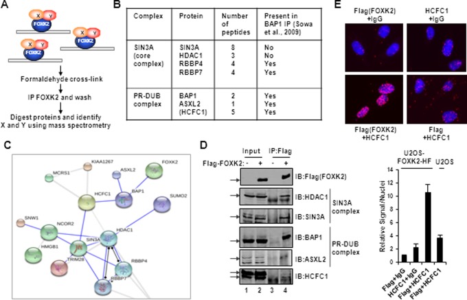
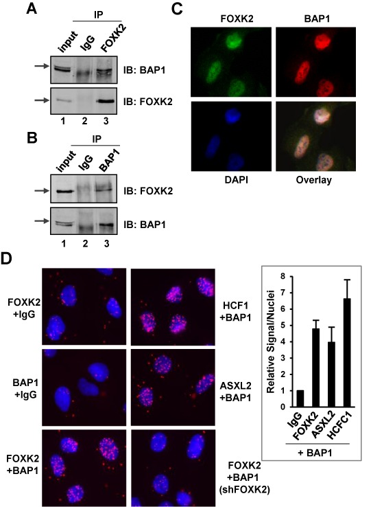
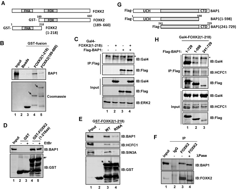
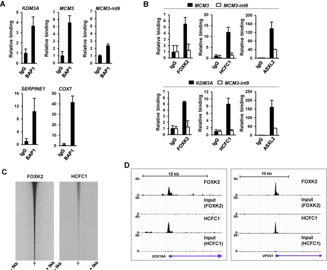
There are numerous forkhead transcription factors in mammalian cells but we know little about the molecular functions of the majority of these. FOXK2 is a ubiquitously expressed family member suggesting an important function across multiple cell types. Here, we show that FOXK2 binds to the SIN3A and PR-DUB complexes. The PR-DUB complex contains the important tumour suppressor protein, the deubiquitinase BAP1. FOXK2 recruits BAP1 to DNA, promotes local histone deubiquitination and causes changes in target gene activity. Our results therefore provide an important link between BAP1 and the transcription factor FOXK2 and demonstrate how BAP1 can be recruited to specific regulatory loci.


Figures

Figure 1.
Figure 1.
Identification of FOXK2 binding proteins by RIME. (A) Schematic illustration of the RIME protocol used to identify FOXK2 associated factors in the context of chromatin association. (B) Summary of FOXK2 interaction proteins belong to the SIN3A core complex and the PR-DUB complex. HCFC1 is shown in brackets as it is unclear whether this is part of the core PR-DUB complex. (C) Visualization using STRING of a sub-network of interactions between FOXK2 binding proteins. (D) Validation of FOXK2 interaction with endogenous components of SIN3A and PR-DUB core complex proteins by co-immunoprecipitation (IP) experiments using anti-Flag (FOXK2) antibody in U2OS–FOXK2–HF cells. Precipitated proteins were detected by immunoblotting (IB) using the antibodies as indicated. Arrows represent the bands corresponding to each of the full-length proteins. Note that the ASXL2 blot is from a different IP experiment. (E) PLA analysis of interaction between Flag-tagged FOXK2 and endogenous HCFC1 in U2OS–FOXK2–HF (top and bottom left panels) or U2OS (bottom right) cells. The combinations of antibodies used are shown above and below each panel (IgG represents a non-specific antibody). DNA is stained using DAPI (blue) and the PLA signal is red. Quantitative analysis of PLA signals in the nucleus is shown below. The level of signals/nuclei in the control PLA sample (Flag and non-specific IgG antibodies) was set as 1.
Figure 2.
Figure 2.
Interactions between endogenous FOXK2 and BAP1. Reciprocal co-immunoprecipitation experiments using either FOXK2 (A) or BAP1 (B) antibodies for immunoprecipitation (IP) from U2OS cells. Co-precipitated endogenous BAP1 and FOXK2 were detected by immunoblotting (IB). Arrows represent the bands corresponding to each of the full-length proteins. (C) Immunofluorescence detection of endogenous FOXK2 (green) and BAP1 (red) and their co-localization in the nucleus [indicated by DAPI staining of DNA (blue)]. (D) Imaging and quantification of PLA signals generated by the indicated combinations of antibodies (IgG represents a non-specific antibody). DNA is stained using DAPI (blue) and the PLA signal is red. Quantitative analysis of PLA signals in the nucleus is shown on the right. The level of signals/nuclei in the control PLA sample (BAP1 and non-specific IgG antibodies) was set as 1.
Figure 3.
Figure 3.
Mapping the FOXK2–BAP1 interaction domains. (A) Schematic representation of full-length FOXK2 and the GST fusions to the C-terminal (amino acids 1–218) and N-terminal (amino acids 189–660) regions of FOXK2. The positions of the forkhead associated (FHA) and forkhead domains (FOX) are shaded in grey. (B) GST pulldown assays using bacterially expressed GST-tagged FOXK2(1–218) and FOXK2(189–660) and in vitro translated BAP1. Arrows mark the positions of full-length GST–FOXK2 fusion proteins. A total of 10% input is shown. (C) Co-immunoprecipitation experiments to analyse interactions between FOXK2(1–218) and BAP1. HEK293T cells were transfected with the indicated plasmids encoding FOXK2(1–218) fused with the Gal4 DNA binding domain and Flag-tagged BAP1, followed by immunoprecipitation (IP) with anti-Flag antibody and immunoblotting (IB) with either an anti-Flag or an anti-Gal4 antibody. ERK2 is a loading control. The asterisk marks a non-specific signal. (D and E) GST pulldown assay using either GST, or wild-type (WT) or FHA mutant (R58A) versions of the GST–FOXK2(1–218) fusion protein and total cell extracts from U2OS cells. Interacting BAP1, HCFC1, SIN3A and input GST fusion proteins were revealed by IB. Arrows mark the positions of full-length GST–FOXK2 fusion proteins. A total of 3% cell lysate input is shown. Ethidium bromide was added to the GST pulldown reactions where indicated. (F) Co-immunoprecipitation experiments using FOXK2 antibodies for IP from U2OS cells. Co-precipitated endogenous BAP1 was detected by IB. Where indicated, the final co-IP was treated with λ phosphatase. (G) Schematic representation of full-length BAP1 and the indicated N- and C-terminal truncation mutants. The locations of the ubiquitin carboxyl-terminal hydrolase (UCH) domain and the C-terminal domain (CTD) are shown. (H) Co-immunoprecipitation analysis of FOXK2 interactions with BAP1 deletion mutants. HEK293T cells were transfected with the indicated plasmids encoding FOXK2(1–218) fused to the Gal4 DNA binding domain and Flag-tagged full-length or truncated mutants of BAP1, followed by IP with anti-Flag antibody and IB with the anti-Gal4, anti-HCFC1 and anti-Flag antibodies. The asterisk marks a non-specific signal.
Figure 4.
Figure 4.
FOXK2 and the PR-DUB bind to the same chromatin regions. ChIP-qPCR analysis of endogenous BAP1 (A) and the other PR-DUB components HCFC1 and ASXL2 (B) binding to the indicated FOXK2 genomic binding regions. Blacks bars in (B) represent binding to FOXK2 binding regions associated with the MCM3 and KDM3A loci; white bars are negative control region which does not bind to FOXK2 (MCM3-int9). Data are normalized against input DNA and shown relative to binding to non-specific IgG (taken as 1). Data are the average of three independent experiments. (C) Genome wide correlation between FOXK2 and HCFC1 (23) binding regions. Heatmap of HCFC1 and FOXK2 read densities mapped onto FOXK2 peak summits and ranked according to FOXK2 signal. (D) UCSC genome browser view of FOXK2 and HCFC1 binding profiles associated with the DDX19A and VPS51 loci.
Figure 5.
Figure 5.
FOXK2 and BAP1 regulate a common set of target genes. (A) Venn diagrams showing the overlap genes which are commonly down-regulated (top) or up-regulated (bottom) following depletion of FOXK2 (12) or BAP1 (22) in U2OS cells. (B) Comparison of the numbers of overlapping genes showing up- or down-regulation following depletion of FOXK2 or BAP1 (blue bars) compared to the numbers expected by chance (grey bars). P-values are shown above the columns. (C) Validation of FOXK2 and BAP1 co-regulated genes. FOXK2 or BAP1 was depleted in U2OS cells and the indicated target gene expression was detected by RT-qPCR. Non-targeting (NT) siRNAs were used as a control. Data are shown relative to the expression seen with NT siRNA (taken as 1) and are the averages plus standard deviations (error bars) from three independent experiments.
Figure 6.
Figure 6.
FOXK2 acts as a chromatin targeting factor for the BAP1-containing deubiquitinase complex. ChIP analysis of endogenous FOXK2 and BAP1 binding to the indicated genomic loci in U2OS cells treated with non-targeting control siRNAs (siNT, black bars) or siRNA against FOXK2 (siFOXK2, white bars) (A) or siRNA against BAP1 (siBAP1, grey bars) (B). ChIP was performed with nonspecific IgG or anti-FOXK2 and BAP1 antibodies; the data are shown relative to the input DNA and are the averages of two (B) or three (A) experiments. (C) ChIP analysis of H2A and H2AK119 ubiquitin levels at the indicated genomic loci in U2OS cells treated with non-targetting control siRNAs (siNT; light grey bars), siRNA against BAP1 (siBAP1; dark grey bars), siRNA against FOXK2 (siFOXK2; white bars) or both BAP1 and FOXK2 (black bars). ChIP was performed with non-specific IgG, anti-H2A and ubiquitinated K119 H2A antibodies (H2Aub); the data are the averages of three experiments. Statistically significant differences between siNT and siBAP1/siFOXK2 treated samples are shown; P-value <0.05 (*) and <0.01(**). (D) Model depicting the role of FOXK2 in nucleating the recruitment of chromatin remodelling complexes to chromatin. FOXK2 recruits both the PR-DUB and the SIN3A complex, potentially through direct or indirect interactions with the shared subunit HCFC1. Question marks denote uncertainty about which interactions are direct. BAP1 can then cause local histone deubiquitination, and histone deacetylation can potentially be achieved through the HDAC1 component of the SIN3A complex.

Similar articles

Cited by

References

    1. Carlsson P., Mahlapuu M. Forkhead transcription factors: key players in development and metabolism. Dev. Biol. 2002;250:1–23. - PubMed
    1. Hannenhalli S., Kaestner K.H. The evolution of Fox genes and their role in development and disease. Nat. Rev. Genet. 2009;10:233–240. - PMC - PubMed
    1. Pierrou S., Hellqvist M., Samuelsson L., Enerbäck S., Carlsson P. Cloning and characterization of seven human forkhead proteins: binding site specificity and DNA bending. EMBO J. 1994;13:5002–5012. - PMC - PubMed
    1. Jolma A., Yan J., Whitington T., Toivonen J., Nitta K.R., Rastas P., Morgunova E., Enge M., Taipale M., Wei G., et al. DNA-binding specificities of human transcription factors. Cell. 2013;152:327–339. - PubMed
    1. Garry D.J., Meeson A., Elterman J., Zhao Y., Yang P., Bassel-Duby R., Williams R.S. Myogenic stem cell function is impaired in mice lacking the forkhead/winged helix protein MNF. Proc. Natl. Acad. Sci. U.S.A. 2000;97:5416–5421. - PMC - PubMed

Publication types

MeSH terms