. 2014 Jun;42(10):6742-52.
doi: 10.1093/nar/gku277. Epub 2014 Apr 19.

The crystal structure of the Split End protein SHARP adds a new layer of complexity to proteins containing RNA recognition motifs

Affiliations

The crystal structure of the Split End protein SHARP adds a new layer of complexity to proteins containing RNA recognition motifs

Fabiana Arieti et al. Nucleic Acids Res. 2014 Jun.

Abstract

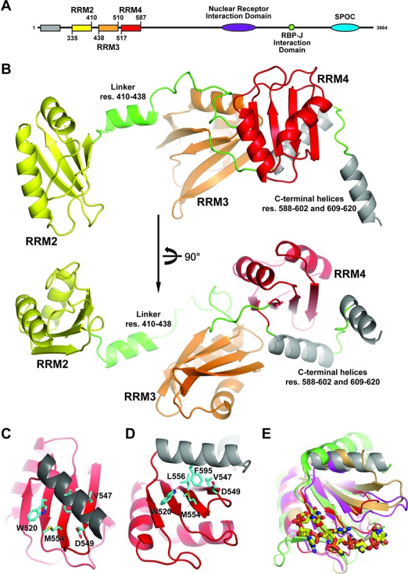
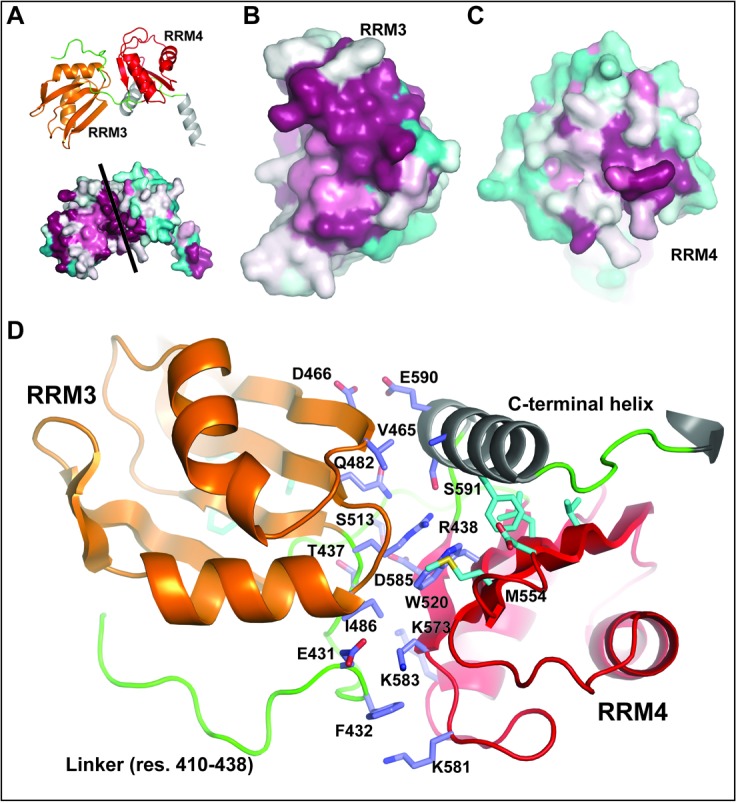
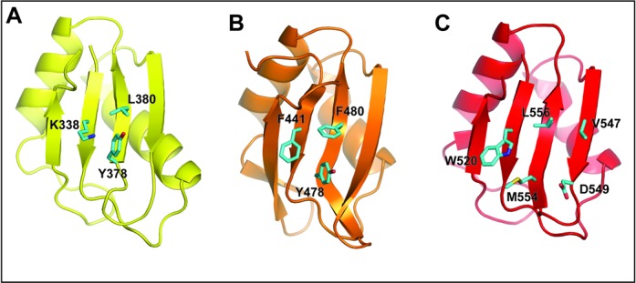
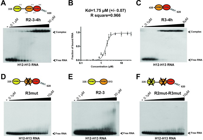
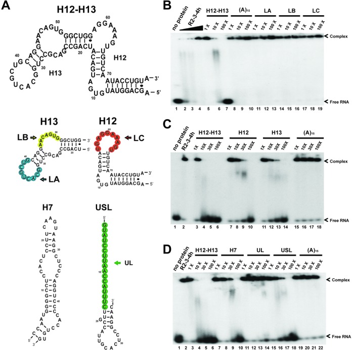
The Split Ends (SPEN) protein was originally discovered in Drosophila in the late 1990s. Since then, homologous proteins have been identified in eukaryotic species ranging from plants to humans. Every family member contains three predicted RNA recognition motifs (RRMs) in the N-terminal region of the protein. We have determined the crystal structure of the region of the human SPEN homolog that contains these RRMs-the SMRT/HDAC1 Associated Repressor Protein (SHARP), at 2.0 Å resolution. SHARP is a co-regulator of the nuclear receptors. We demonstrate that two of the three RRMs, namely RRM3 and RRM4, interact via a highly conserved interface. Furthermore, we show that the RRM3-RRM4 block is the main platform mediating the stable association with the H12-H13 substructure found in the steroid receptor RNA activator (SRA), a long, non-coding RNA previously shown to play a crucial role in nuclear receptor transcriptional regulation. We determine that SHARP association with SRA relies on both single- and double-stranded RNA sequences. The crystal structure of the SHARP-RRM fragment, together with the associated RNA-binding studies, extend the repertoire of nucleic acid binding properties of RRM domains suggesting a new hypothesis for a better understanding of SPEN protein functions.


Figures

Figure 1.
Figure 1.
Overall structure of SHARP–RRMs. (A) Schematic representation of the domain organization of the SHARP protein. (B) The overall structure of R2–3–4h is shown: RRM2 is coloured in yellow, RRM3 in orange, RRM4 in red, C-terminal helices in grey, and linker regions are coloured in green. (C) and (D) Top (C) and side (D) views of the RRM4 plus the C-terminal α-helix. The protein is shown as a cartoon and coloured in red for the RRM and grey for the helix. (E) Superposition of various RRMs followed by C-terminal helices. The xRRM found in the p65 protein is coloured in green (PDB code 4EYT). The qRRM 1 and 2 from the hnRNP F in complex with the AGGGAU hexa-ribonucleotide are coloured in gold and magenta, respectively. The nucleotides are colored according to atom types (carbon: yellow; nitrogen: blue; oxygen: red; and phosphate: orange). SHARP is coloured in red with the helix coloured in grey. Proteins are shown as a cartoon and residues involved in intramolecular interactions in panels C and D are shown as sticks and coloured according to atom type (carbon: cyan; nitrogen: blue; and oxygen: red).
Figure 2.
Figure 2.
RRM3–RRM4 interface conservation. (A) RRM3 and RRM4 (shown as cartoon and coloured in orange and red, respectively) are in close contact. Bottom part shows the same protein fragment using a surface representation coloured according to surface conservation (cyan: low conservation and magenta: high conservation). (B) and (C) The RRM3–RRM4 interface is facing the reader, and the protein surface is coloured as in panel A. (D) Magnified view of the RRM3–RRM4 interface of SHARP: residues located at the interface are shown as sticks and coloured according to atom types (carbon: slate or cyan; nitrogen: blue; and oxygen: red).
Figure 3.
Figure 3.
Individual RRM structures and the consensus amino acids for RNA binding. (A), (B), and (C) RRM2, RRM3, and RRM4 are shown as a cartoon and coloured in yellow, orange, and red, respectively. Amino acids at the canonical position for RNA interaction are shown as sticks and coloured as in Figure 1.
Figure 4.
Figure 4.
Identification of the protein region responsible for the SRA RNA association. (A) Representative EMSA used to quantify R2–3–4h construct association with the SRA RNA fragment H12–H13. (B) Quantification of the EMSA experiment shown in panel A. The bound fraction was quantified and analysed using the Hill Equation, and the fit is shown as a solid line. The calculated dissociation constant (Kd, app) and the quality of the fit are indicated. (C) EMSA autoradiogram obtained when measuring the RNA-binding capacity of the R3-R4h construct. (D) EMSA autoradiogram showing the very weak RNA-binding activity of the R3mut construct. (E) EMSA autoradiogram showing the poor RNA-binding activity of the R2–3 construct. (F) EMSA autoradiogram obtained using the R2mutR3mut construct and indicating that the RRM4 plus the two helices participate only marginally in RNA association.
Figure 5.
Figure 5.
SHARP–RRM association with RNA relies on stem-loop structure. (A) Secondary structures and sequences of the RNA molecules used for the competition assays reported in panels B, C, and D. The structural architecture of the H12–H13 fragment is based on that established by Novikova et al.(38). The loop regions and the single-strand RNA are coloured in blue (LA), yellow (LB), red (LC), and green (UL). (B) RNA-binding competition assays to determine the loop sequences mediating association between SHARP–RRM (R2–3–4h construct) and the SRA RNA H12–H13. Free RNA and R2–3–4h complex are shown in lanes 1 and 2. Competition assays were performed with the following RNAs: loop LA (lanes 11–13), loop LB (lanes 14–16), and loop LC (lanes 17–19). (C) Similar competition experiments performed with individual substructures H12 and H13 from the SRA RNA. Competition assays were performed with H12 RNA (lanes 7–10) and H13 RNA (lanes 11–14). (D) Competition experiments performed with various unrelated RNAs showing the importance of a stem region for the competitive capacity of the RNA. Competition assays were performed with cold H7 RNA (lanes 7–10), unrelated loop RNA (UL, lanes 11–14), and unrelated stem-loop RNA (USL, lanes 15–18). Concentrations of the cold RNAs were added in molar excess as indicated in each panel. Positive and negative controls for efficient competitions are identical in panels B, C, and D (labelled H12–H13 or A15, respectively).

Similar articles

Cited by

References

    1. Dickson B.J., van der Straten A., Dominguez M., Hafen E. Mutations modulating Raf signaling in Drosophila eye development. Genetics. 1996;142:163–171. - PMC - PubMed
    1. Kolodziej P.A., Jan L.Y., Jan Y.N. Mutations that affect the length, fasciculation, or ventral orientation of specific sensory axons in the Drosophila embryo. Neuron. 1995;15:273–286. - PubMed
    1. Wiellette E.L., Harding K.W., Mace K.A., Ronshaugen M.R., Wang F.Y., McGinnis W. Spen encodes an RNP motif protein that interacts with Hox pathways to repress the development of head-like sclerites in the Drosophila trunk. Development. 1999;126:5373–5385. - PubMed
    1. Yabe D., Fukuda H., Aoki M., Yamada S., Takebayashi S., Shinkura R., Yamamoto N., Honjo T. Generation of a conditional knockout allele for mammalian Spen protein Mint/SHARP. Genesis. 2007;45:300–306. - PubMed
    1. Newberry E.P., Latifi T., Towler D.A. The RRM domain of MINT, a novel Msx2 binding protein, recognizes and regulates the rat osteocalcin promoter. Biochemistry. 1999;38:10678–10690. - PubMed

Publication types

Associated data