

doi: 10.1016/j.eats.2013.09.007.
eCollection 2014 Feb.
Endoscopic transtendinous repair for partial-thickness proximal hamstring tendon tears
Affiliations
- PMID: 24749032
- PMCID: PMC3986502
- DOI: 10.1016/j.eats.2013.09.007
Item in Clipboard
Endoscopic transtendinous repair for partial-thickness proximal hamstring tendon tears
Arthrosc Tech.
.
Abstract
Partial tears of the proximal hamstring tendon can successfully be managed with tendon repair in cases of failed conservative management. As in partial-thickness gluteus medius repair, a transtendinous technique can be used to repair partial-thickness undersurface tears of the hamstring origin. This report details an endoscopic transtendinous approach for the treatment of partial-thickness hamstring tendon tears.
Figures
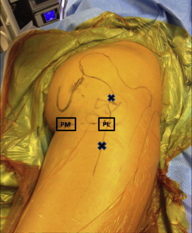
The patient is positioned prone with the entire right lower extremity draped free. The posteromedial (PM) and posterolateral (PL) portals are marked. Access to the ischium is obtained first through the posterolateral portal under fluoroscopic guidance. Accessory portals (X) are used for suture passage and management as necessary.
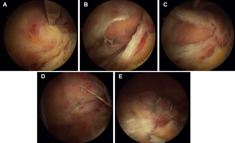
(A) Endoscopic photograph of right hamstring insertion, viewing from posteromedial portal with 70° arthroscope. The incision is made into the hamstring insertion with a beaver blade. (B) Elevation of lateral edge of hamstring insertion. (C) Elevation of medial edge of hamstring insertion. A burr is used to decorticate the bone underlying the free edges. (D) After passage of 2 anchors and all sutures, tension on the sutures reapproximates the free ends. (E) Final repair with secure fixation of hamstring insertion to ischial tuberosity.

(A) Coronal T2-weighted image with fat saturation showing the common insertion of the hamstring tendons (white arrow) into the ischial tuberosity. One should note the clear separation, noted by fluid (red arrow), between the tendons and the ischium (plus sign). (B) Axial T2-weighted image with fat saturation showing separation of hamstring from its insertion. The red line indicates the location and angle of approach for the beaver blade to make a longitudinal cut in the partial hamstring tendon tear.
References
-
- Wood D.G., Packham I., Trikha S.P., Linklater J. Avulsion of the proximal hamstring origin. J Bone Joint Surg Am. 2008;90:2365–2374. - PubMed
-
- Aldridge S.E., Heilpern G.N., Carmichael J.R., Sprowson A.P., Wood D.G. Incomplete avulsion of the proximal insertion of the hamstring: Outcome two years following surgical repair. J Bone Joint Surg Br. 2012;94:660–662. - PubMed
-
- Bowman K.F., Jr., Cohen S.B., Bradley J.P. Operative management of partial-thickness tears of the proximal hamstring muscles in athletes. Am J Sports Med. 2013;41:1363–1371. - PubMed
-
- Domb B.G., Nasser R.M., Botser I.B. Partial-thickness tears of the gluteus medius: Rationale and technique for trans-tendinous endoscopic repair. Arthroscopy. 2010;26:1697–1705. - PubMed
LinkOut - more resources
Full Text Sources
Other Literature Sources
Research Materials
