. 2014 Apr 3;2(4):406-13.
doi: 10.1016/j.stemcr.2014.02.008. eCollection 2014 Apr 8.

Do neonatal mouse hearts regenerate following heart apex resection?

Affiliations

Do neonatal mouse hearts regenerate following heart apex resection?

Ditte Caroline Andersen et al. Stem Cell Reports. .

Abstract

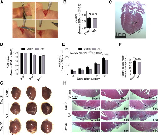
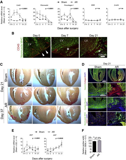
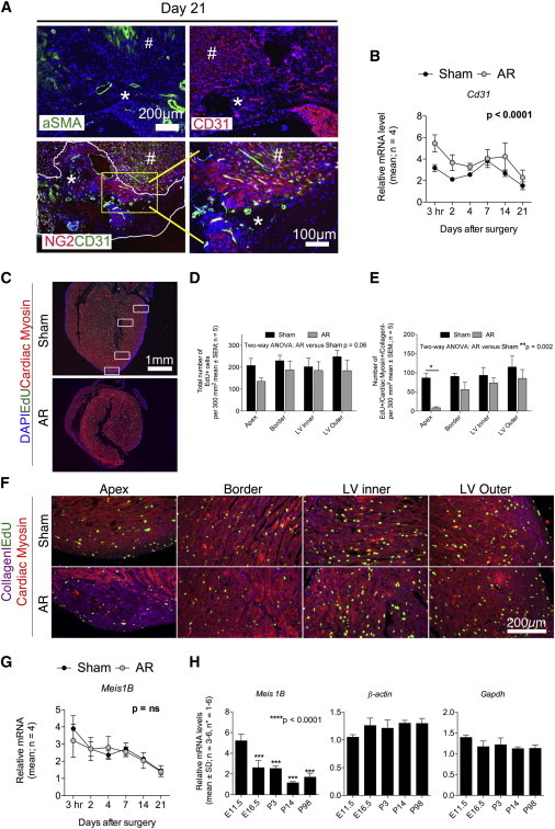
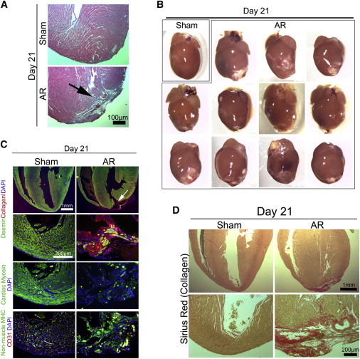
The mammalian heart has generally been considered nonregenerative, but recent progress suggests that neonatal mouse hearts have a genuine capacity to regenerate following apex resection (AR). However, in this study, we performed AR or sham surgery on 400 neonatal mice from inbred and outbred strains and found no evidence of complete regeneration. Ideally, new functional cardiomyocytes, endothelial cells, and vascular smooth muscle cells should be formed in the necrotic area of the damaged heart. Here, damaged hearts were 9.8% shorter and weighed 14% less than sham controls. In addition, the resection border contained a massive fibrotic scar mainly composed of nonmyocytes and collagen disposition. Furthermore, there was a substantial reduction in the number of proliferating cardiomyocytes in AR hearts. Our results thus question the usefulness of the AR model for identifying molecular mechanisms underlying regeneration of the adult heart after damage.


Figures

Figure 1
Figure 1
C57Bl/6 Neonatal Mouse Hearts Do Not Regenerate following AR (A) Open thoracotomy was used to remove 5%–10% of the heart apex from hypothermia-anesthetized neonatal C57Bl/6 mice. (B and C) Hearts from sham (n = 21) and apex-resected (n = 23) mice were removed immediately after AR and weighed to determine the amount of resected heart tissue. Hematoxylin staining of AR hearts (n = 10) confirmed localization of the resected area. Black line indicates resection border. LV, left ventricle. (D) Animal survival was determined immediately (0 hr), then 3 and 24 hr after surgery. (E) The cardiac weight in sham and AR animals was determined at different time points following surgery. Two-way ANOVA was used to test significance (∗∗∗∗p < 0.0001 for weight over time). (F) Relative cardiac height at day 21 in sham (n = 5) and AR (n = 7) hearts. Student’s t test was used to test significance (p < 0.05). (G) Three representative stereomicroscopic pictures of sham and AR hearts. (H) Representative HE stainings from three to six sham or AR hearts at day 21 or 37 that enclose the AR damage. All hearts (n = 16) were sectioned throughout and examined (see Figure S1) to identify the area of apex lesion marked by an asterisk (). See also Figure S1.
Figure 2
Figure 2
C57Bl/6 Hearts Become Substantially Fibrotic following AR and Show Signs of Hypertrophy (A) Quantitative real-time PCR of sham and AR hearts (n = 4) at indicated time points. Statistical significance tested by two-way ANOVA is indicated. (B and D) Paraffin- and cryo-embedded C57Bl/6 AR hearts (n = 4–8) were sectioned and immunostained for (B) CD45/nonmuscle myosin and (D) desmin/collagen; cardiac myosin; nonmuscle myosin/CD31. Representative images were processed (contrast/brightness and color balance) equally in Adobe Photoshop to enable merging. An asterisk () indicates lesion area, whereas white arrows reflect the AR line. (C) Sirius Red stainings of representative sham (n = 5) and AR (n = 11) heart sections from C57Bl/6 mice identify collagen disposition in the lesioned apex. (E) Quantitative real-time PCR for hypertrophic markers (ANP and BNP) of sham and AR hearts (n = 4) at indicated time points. (F) The relative width of C57Bl/6 hearts from sham and AR animals was measured at day 21. Significance was tested by a Student’s t test (p < 0.05). For (A) and (E), quantitative real-time PCR raw data were normalized against B2M and β-actin, which were stably expressed as determined by the qBase+ platform (M:0.579 and CV:0.202) (Hellemans et al., 2007, Vandesompele et al., 2002). See Figure S2 for quantitative real-time PCR profiling on gene programs at day 21 following AR.
Figure 3
Figure 3
The Apex-Resected Zone in C57Bl/6 Hearts Reveals Limited Vascularization and Numbers of Proliferating Cardiomyocytes (A) Cryosectioned C57Bl/6 AR hearts (n = 4–8) were immunostained for aSMA, CD31, and NG2/CD31. Representative images were processed (contrast/brightness and color balance) equally in Photoshop to enable merging. An asterisk () indicates lesion area, whereas a number sign (#) refers to nondamaged myocardium. (B) Quantitative real-time PCR for Cd31 of sham and AR hearts (n=4) at indicated time points. Quantitative real-time PCR raw data were normalized against the stably expressed B2M and β-actin genes (see Figure 2). (C–F) Paraffin-embedded C57Bl/6 AR and sham hearts (n = 5) from EdU pulse-chase labeled animals were sectioned and immunostained for EdU/cardiac myosin/collagen I/DAPI to enable identification of proliferating cells in total and proliferating cardiomyocytes specifically. Images were taken in four different zones (white boxes in C and areas magnified in F) of each heart and used for cell number quantification (D and E) as described in Experimental Procedures. (G) Quantitative real-time PCR for Meis1b of sham and AR hearts (n = 4) at indicated time points. Quantitative real-time PCR raw data were normalized against the stably expressed B2M and β-actin genes (see Figure 2). (H) Meis1b is downregulated with cardiac development. Hearts were microdissected from C57Bl/6 mice at indicated time points and used for quantitative real-time PCR. Primers used were previously described by Mahmoud et al. (2013). Each biological experiment (n = 3–6) included between one and six hearts. qPCR raw data were normalized against Gapdh and β-actin, which were stably expressed as determined by the qBase+ platform (M:0.306, CV: 0.106) (Hellemans et al., 2007, Vandesompele et al., 2002). Statistical significance was tested using a one-way ANOVA with a Dunnett’s multiple comparison posttest. E11.5, embryonic day 11.5.
Figure 4
Figure 4
Apex-Resected ICR/CD1 Hearts Do Not Reveal Any Major Regenerative Response but Become Fibrotic Representative sham (n = 3) and AR (n = 11) hearts from outbred ICR/CD-1 neonates (n = 24) were analyzed at day 21 by (A) HE staining (arrow identifies lesion), (B) stereomicroscopy, (C) immunofluorescence, and (D) Sirius Red staining. Representative sections are shown (see Figure S3). For (C), images were processed (contrast/brightness and color balance) equally in Photoshop to enable merging.

Comment in

References

    1. Aguirre A., Sancho-Martinez I., Izpisua Belmonte J.C. Reprogramming toward heart regeneration: stem cells and beyond. Cell Stem Cell. 2013;12:275–284. - PubMed
    1. Aldinger K.A., Sokoloff G., Rosenberg D.M., Palmer A.A., Millen K.J. Genetic variation and population substructure in outbred CD-1 mice: implications for genome-wide association studies. PLoS ONE. 2009;4:e4729. - PMC - PubMed
    1. Andersen D.C., Petersson S.J., Jørgensen L.H., Bollen P., Jensen P.B., Teisner B., Schroeder H.D., Jensen C.H. Characterization of DLK1+ cells emerging during skeletal muscle remodeling in response to myositis, myopathies, and acute injury. Stem Cells. 2009;27:898–908. - PubMed
    1. Ang K.L., Shenje L.T., Reuter S., Soonpaa M.H., Rubart M., Field L.J., Galiñanes M. Limitations of conventional approaches to identify myocyte nuclei in histologic sections of the heart. Am. J. Physiol. Cell Physiol. 2010;298:C1603–C1609. - PMC - PubMed
    1. Drenckhahn J.D., Schwarz Q.P., Gray S., Laskowski A., Kiriazis H., Ming Z., Harvey R.P., Du X.J., Thorburn D.R., Cox T.C. Compensatory growth of healthy cardiac cells in the presence of diseased cells restores tissue homeostasis during heart development. Dev. Cell. 2008;15:521–533. - PubMed

Publication types