

Neural stem cells and glioblastoma
- PMID: 24750704
- PMCID: PMC4202863
- DOI: 10.15274/NRJ-2014-10028
Neural stem cells and glioblastoma
Abstract
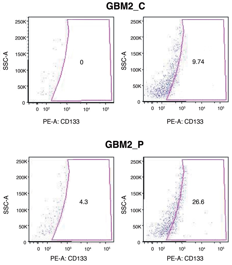
Glioblastoma multiforme represents one of the most common brain cancers with a rather heterogeneous cellular composition, as indicated by the term "multiforme". Recent reports have described the isolation and identification of cancer neural stem cells from human adult glioblastoma multiforme, which possess the capacity to establish, sustain, and expand these tumours, even under the challenging settings posed by serial transplantation experiments. Our study focused on the distribution of neural cancer stem cells inside the tumour. The study is divided into three phases: removal of tumoral specimens in different areas of the tumour (centre, periphery, marginal zone) in an operative room equipped with a 1.5 T scanner; isolation and characterization of neural cancer stem cells from human adult glioblastoma multiforme; identification of neural cancer stem cell distribution inside the tumour.
Keywords: brainsuite; glioblastoma; neural stem cells.
Figures


References
-
- Jellinger K. Glioblastoma multiforme: morphology and biology. Acta Neurochir (Wien) 1978;42(1-2):5–32. doi: 10.1007/BF01406628. - DOI - PubMed
-
- Galli R, Binda E, Orfanelli U, et al. Isolation and characterization of tumorigenic, stem-like neural precursors from human glioblastoma. Cancer Res. 2004;64(19):7011–7021. doi: 10.1158/0008-5472.CAN-04-1364. - DOI - PubMed
-
- Beier D, Hau P, Proescholdt M, et al. CD133(+) and CD133(-) glioblastoma-derived cancer stem cells show differential growth characteristics and molecular profiles. Cancer Res. 2007;67(9):4010–4015. doi: 10.1158/0008-5472.CAN-06-4180. - DOI - PubMed
-
- Vescovi A. Brain tumour stem cells. Nat Rev Cancer. 2006;6(6):425–436. doi: 10.1038/nrc1889. - DOI - PubMed
-
- Pagano SF, Impagnatiello F, Girelli M, et al. Isolation and characterization of neural stem cells from the adult human olfactory bulb. Stem Cells. 2000;18:295–300. doi: 10.1634/stemcells.18-4-295. - DOI - PubMed
MeSH terms
Substances
LinkOut - more resources
Full Text Sources
Other Literature Sources
Medical
