. 2014 Dec;15(9):881-91.
doi: 10.1111/mpp.12146. Epub 2014 Jun 5.

Patellins 3 and 6, two members of the Plant Patellin family, interact with the movement protein of Alfalfa mosaic virus and interfere with viral movement

Affiliations

Patellins 3 and 6, two members of the Plant Patellin family, interact with the movement protein of Alfalfa mosaic virus and interfere with viral movement

Ana Peiro et al. Mol Plant Pathol. 2014 Dec.

Abstract

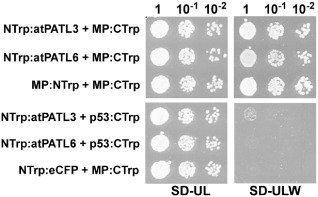
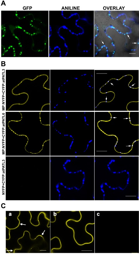
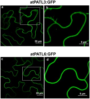
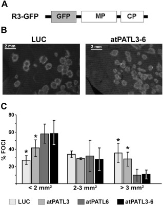
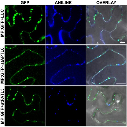
Movement proteins (MPs) encoded by plant viruses interact with host proteins to facilitate or interfere with intra- and/or intercellular viral movement. Using yeast two-hybrid and bimolecular fluorescence complementation assays, we herein present in vivo evidence for the interaction between Alfalfa mosaic virus (AMV) MP and Arabidopsis Patellin 3 (atPATL3) and Patellin 6 (atPATL6), two proteins containing a Sec14 domain. Proteins with Sec14 domains are implicated in membrane trafficking, cytoskeleton dynamics, lipid metabolism and lipid-mediated regulatory functions. Interestingly, the overexpression of atPATL3 and/or atPATL6 interfered with the plasmodesmata targeting of AMV MP and correlated with reduced infection foci size. Consistently, the viral RNA levels increased in the single and double Arabidopsis knockout mutants for atPATL3 and atPATL6. Our results indicate that, in general, MP-PATL interactions interfere with the correct subcellular targeting of MP, thus rendering the intracellular transport of viral MP-containing complexes less efficient and diminishing cell-to-cell movement.

Keywords: AMV; ilarvirus; intercellular movement; movement protein; patellin.


Figures

Figure 1
Figure 1
Trp1 yeast two‐hybrid assays. Different dilutions (top) of yeast cells co‐transformed with the indicated pair of plasmids (left) were spotted onto synthetic medium containing (SD‐UL) or lacking (SD‐ULW) tryptophan to confirm the correct transformation or positive interactions, respectively. Self‐interaction of the movement protein (MP) (MP:NTrp + MP:CTrp) was used as a positive interaction. Cells co‐transformed with NTrp:atPATL3 or NTrp:atPATL6 plus p53:CTrp or MP:CTrp plus NTrp:eCFP were used as negative controls.
Figure 2
Figure 2
Localization of Alfalfa mosaic virus (AMV) movement protein (MP) at plasmodesmata (PD) and bimolecular fluorescence complementation (BiFC) analysis of the MPPatellin (MPPATL) interactions. (A) Confocal laser scanning microscopy (CLSM) images of epidermal cells expressing MP:GFP [green fluorescent protein (GFP) panel] and stained with aniline blue (ANILINE panel) showing MP:GFP and callose localization, respectively. OVERLAY panel is the superposition of GFP, ANILINE and the corresponding bright field image. Arrows indicate PD labelled with both MP:GFP and aniline blue. (B) BiFC analysis to corroborate AMV MP–atPATL interaction in planta. CLSM images of epidermal cells co‐infiltrated with MP:NYFP and CYFP:atPATL3 or CYFP:atPATL6 (indicated on the left) and stained with aniline blue solution. Panels on the right are the superposition of the yellow fluorescent protein (YFP) fluorescence and Aniline staining images (panels on the left and centre, respectively). Arrows indicate reconstituted fluorescence co‐localizing with callose‐rich PD. Leaves infiltrated with NYFP and CYFP:atPATL3 are the negative interaction controls. (C) BiFC analysis to analyse the implication of the GOLD domain in the interaction between AMV MP and atPATL3. CLSM images of epidermal leaves co‐infiltrated with MP:NYFP and CYFP:atPATL3 (a), MP:NYFP and CYFP:atPATLP3‐ΔGOLD (b) or MP:NYFP and CYFP:GOLD‐P3 (c) are shown. Arrows indicate fluorescence spots representing PD. Bar, 10 μm.
Figure 3
Figure 3
Subcellular localization of Arabidopsis Patellin3 (atPATL3) and atPATL6. Confocal laser scanning microscopy (CLSM) images of epidermal cells infiltrated with AgrobacteriumC58 expressing atPATL3 and atPATL6 with the green fluorescent protein (GFP) fused at their C‐terminus (atPATL3:GFP and atPATL6:GFP, respectively). Both fusion proteins present a strong signal at the cell periphery. (b) and (d) show enlarged images of the boxed areas.
Figure 4
Figure 4
Effect of Arabidopsis Patellin (atPATL) over‐expression on the viral infection. (A) Schematic representation showing the modified Alfalfa mosaic virus (AMV) RNA 3 expressing the green fluorescent protein (GFP) used in this study (R3‐GFP). The open reading frames corresponding to GFP, movement protein (MP) and coat protein (CP) are shown as boxes. (B) Representative images of the foci induced by R3‐GFP in leaves infiltrated with Agrobacterium expressing luciferase (LUC) or a mixture of both atPATLs (atPATL3–6). (C) Graphic showing the average percentage of foci grouped into three different categories according to size area in leaves overexpressing LUC, atPATL3, atPATL6 or a mixture of both (atPATL3–6). Standard deviation values are shown. Significant differences are indicated by *P < 0.05.
Figure 5
Figure 5
Viral accumulation in Arabidopsis Patellin (atPATL) knockouts. (A) Image of Arabidopsis wild‐type (wt) and knockout seedlings germinated on Murashige and Skoog medium. (B) Detection of Alfalfa mosaic virus (AMV) RNA 3 and 4 accumulation (indicated on the left) in wt and knockout lines by Northern blot analysis of five infected plants. Bottom panel shows the ethidium bromide (EtBr)‐stained gel as a loading control (it only shows the band corresponding to the 25S ribosomal RNA). (C) Graphic showing the average of viral RNA accumulation measured from the Northern blot in (B). Standard deviation values are shown. Significant differences are indicated by *P < 0.05.
Figure 6
Figure 6
Alfalfa mosaic virus (AMV) movement protein (MP) subcellular localization in the presence of Arabidopsis Patellins (atPATLs). Images of epidermal cells co‐infiltrated with Agrobacterium expressing the proteins indicated on the left and stained with aniline blue. Overlay panels correspond to the superposition of green fluorescent protein (GFP), ANILNE and the corresponding bright field images. Bar, 10 μm.

Similar articles

Cited by

References

    1. Allen‐Baume, V. , Segui, B. and Cockcroft, S. (2002) Current thoughts on the phosphatidylinositol transfer protein family. FEBS Lett. 531, 74–80. - PubMed
    1. Anantharaman, V. and Aravind, L. (2002) The GOLD domain, a novel protein module involved in Golgi function and secretion. Genome Biol. 3, 1–7. - PMC - PubMed
    1. Aparicio, F. , Sánchez‐Navarro, J.A. and Pallas, V. (2006) In vitro and in vivo mapping of the Prunus necrotic ringspot virus coat protein C‐terminal dimerization domain by bimolecular fluorescence complementation. J. Gen. Virol. 87, 1745–1750. - PubMed
    1. Aparicio, F. , Sánchez‐Navarro, J.A. and Pallas, V. (2010) Implication of the C terminus of the Prunus necrotic ringspot virus movement protein in cell‐to‐cell transport and in its interaction with the coat protein. J. Gen. Virol. 91, 1865–1870. - PubMed
    1. Aparicio, F. , Aparicio‐Sanchis, R. , Gadea, J. , Sánchez‐Navarro, J.A. , Pallás, V. and Murguía, J.R. (2011) A plant virus movement protein regulates the Gcn2p kinase in budding yeast. PLoS ONE, 6, e27409. - PMC - PubMed

Publication types

MeSH terms

Substances