

doi: 10.3201/eid2005.130778.
Chronic wasting disease agents in nonhuman primates
- PMID: 24751215
- PMCID: PMC4012792
- DOI: 10.3201/eid2005.130778
Item in Clipboard
Chronic wasting disease agents in nonhuman primates
Emerg Infect Dis.
2014 May.
Abstract
Chronic wasting disease is a prion disease of cervids. Assessment of its zoonotic potential is critical. To evaluate primate susceptibility, we tested monkeys from 2 genera. We found that 100% of intracerebrally inoculated and 92% of orally inoculated squirrel monkeys were susceptible, but cynomolgus macaques were not, suggesting possible low risk for humans.
Keywords: Chronic wasting disease; cynomolgus macaque; non-human primate; prion; species barrier; squirrel monkey; transmissible spongiform encephalopathy; transmission.
Figures
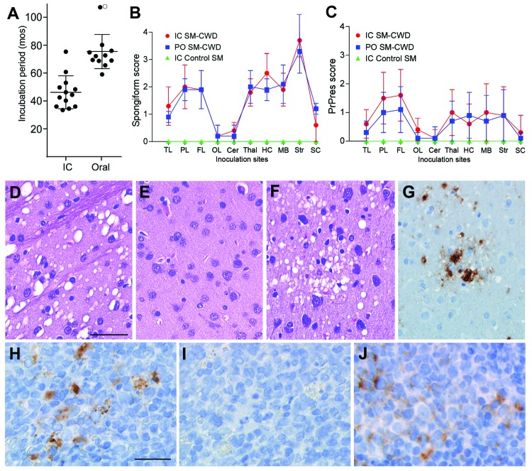
Incubation periods of chronic wasting disease (CWD) and neuropathologic features of CWD agent–infected squirrel monkeys. A) Incubation periods for squirrel monkeys infected with CWD agents by intracerebral (IC) or oral (PO) routes. Solid circles indicate euthanized squirrel monkeys (SM) that tested positive for prion disease. The open circle indicates 1 squirrel monkey that remained clinically normal at 108 months postinoculation (mpi). Lines indicate the mean and standard deviation within each group. B, C) Lesion profiles of CWD-agent–infected squirrel monkeys showing spongiform degeneration (B) and PrPres deposition (C) values in 10 gray matter regions of the brain. N values for each group are as follows: IC SM-CWD, 11; PO SM-CWD, 7; IC control SM, 1. TL, temporal lobe; PL, parietal lobe; FL, frontal lobe; OL, occipital lobe; Cer, cerebellum; Thal, thalamus; HC, hippocampus; MB, midbrain; Str, stiatum; SC, spinal cord. Error bars show the SD for each group. Panels D-G show brain from a squirrel monkey infected PO with CWD and euthanized at 69 months postinoculation. Panels D-F are stained with hematoxylin and eosin and show D) severe spongiform lesions in the striatum, E) lack of pathology in the occipital lobe, and F) pathology in the parietal lobe. Panels G, H, and J show immunohistochemical staining for PrPres by using anti-PrP antibody D13. G) Adjacent section to the region depicted in F shows the positive correlation of PrPres (brown) with spongiform degeneration. Panels H–J show lymphoid tissue from a squirrel monkey infected PO with CWD and euthanized at 80 mpi. H) PrPres (brown) staining in spleen and J) mesenteric lymph node. I) No primary antibody control of the region shown in H, demonstrating specificity of stain observed in H. The scale bar shown in D applies to panels D–G and represents 50 µm; the scale bar shown in H applies to H–I and represents 25 µm.
Neuropathologic features and immunoblot results of second-passage squirrel monkeys that had chronic wasting disease (CWD). Scale bar represents 50 µM and is applicable to panels A and B. Panels A and B show neuropathologic changes in the occipital lobe of SMP2-CWD monkey 977, which was euthanized at 24 months postinoculation. A) Hematoxylin and eosin staining show prominent spongiform changes. B) Immunohistochemical staining for disease-associated prion protein (PrPres) (brown) with anti-PrP antibody D13. C) Results of Western blot for PrPres in brain tissue of cervids and its respective first and second passage in squirrel monkeys. MD-1 was used to infect SM308, and SM308 was used to infect SM977. Lanes 1, 2, 5, and 6, 0.6 mg brain equivalents. Lanes 3 and 7, 0.36 mg brain equivalents to give similar signal intensities to the other samples. Lane 4, blank (Bl). Apparent molecular weights (in kDa) are provided on the left side of panel C. Immunoblot was probed with anti-PrP antibody L42. When comparing the 2 central bands, cervid CWD had a more intense band at 25.5 kDa; SM-CWD (nos. 308 and 322) and SM2-CWD (nos. 977 and 840) were more intense at 27 kDa.
References
Publication types
MeSH terms
Substances
Grants and funding
LinkOut - more resources
Full Text Sources
Other Literature Sources
