. 2014 Apr 21;9(4):e95100.
doi: 10.1371/journal.pone.0095100. eCollection 2014.

Expression of Caveolin 1 is enhanced by DNA demethylation during adipocyte differentiation. status of insulin signaling

Affiliations

Expression of Caveolin 1 is enhanced by DNA demethylation during adipocyte differentiation. status of insulin signaling

Sara Palacios-Ortega et al. PLoS One. .

Abstract

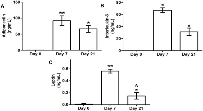
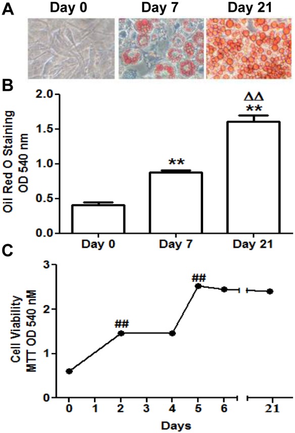
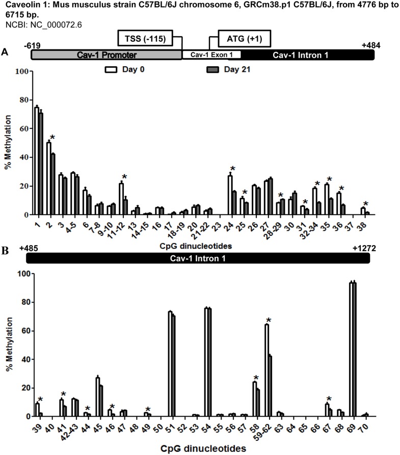
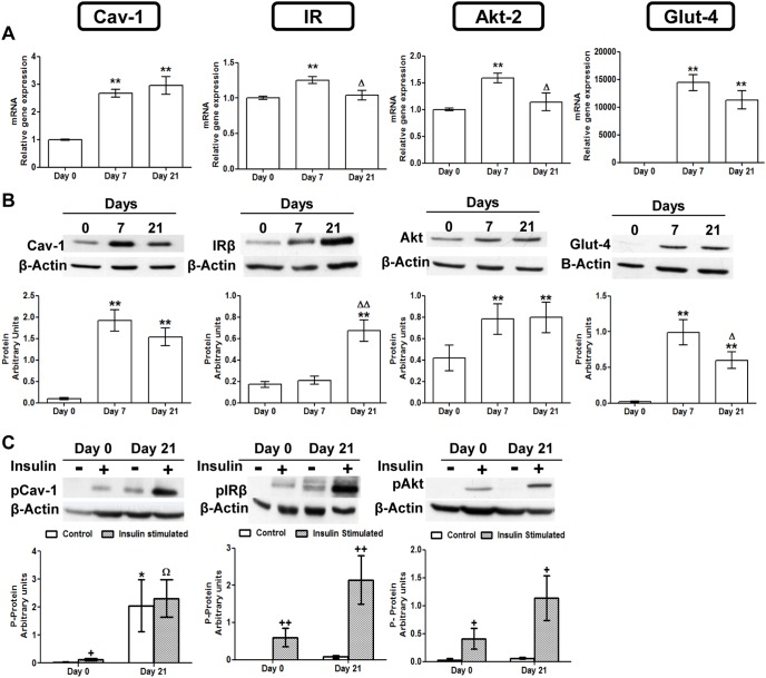
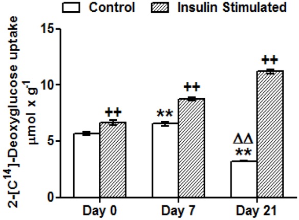
Caveolin 1 (Cav-1) is an essential constituent of adipocyte caveolae which binds the beta subunit of the insulin receptor (IR) and is implicated in the regulation of insulin signaling. We have found that, during adipocyte differentiation of 3T3-L1 cells the promoter, exon 1 and first intron of the Cav-1 gene undergo a demethylation process that is accompanied by a strong induction of Cav-1 expression, indicating that epigenetic mechanisms must have a pivotal role in this differentiation process. Furthermore, IR, PKB-Akt and Glut-4 expression are also increased during the differentiation process suggesting a coordinated regulation with Cav-1. Activation of Cav-1 protein by phosphorylation arises during the differentiation process, yet in fully mature adipocytes insulin is no longer able to significantly increase Cav-1 phosphorylation. However, these long-term differentiated cells are still able to respond adequately to insulin, increasing IR and PKB-Akt phosphorylation and glucose uptake. The activation of Cav-1 during the adipocyte differentiation process could facilitate the maintenance of insulin sensitivity by these fully mature adipocytes isolated from additional external stimuli. However, under the influence of physiological conditions associated to obesity, such as chronic inflammation and hypoxia, insulin sensitivity would finally be compromised.


Conflict of interest statement

Competing Interests: The authors have declared that no competing interests exist.

Figures

Figure 1
Figure 1. Secreted adiponectin interleukin 6 (IL-6) and leptin levels during 3T3-L1 cell adipogenesis.
Culture medium samples of 3T3-L1 preadipocytes (d0) and adipocytes (d7, d21) were obtained for adipokine ELISA assay. Data are means ± SEM of the concentration (ng/mL) of each adipokine secreted to the cell medium. (A) Adiponectin (B) Interleukin 6 (IL-6) (C) Leptin. Groups were compared using the Wilcoxon signed rank test. Data are referred to day 0 *. p≤0.05, **. p≤0.01 or to day 7 Δ. p≤0.05.
Figure 2
Figure 2. 3T3-L1 cell differentiation and viability.
(A) Cells were photographed at days 0, 7 and 21 of their adipocytic differentiation after Oil Red O staining using a light microscope (40X magnification). Data are means ± SEM of dye OD at 540 nm. (B) Intracellular triglycerides quantification at days 0, 7 and 21 using Oil Red O Staining. (C) Cell viability measurement at days 0, 2, 4, 5, 6 and 21 using MTT assay. Data are means ± SEM of dye OD at 540 nm. Groups were compared using the ANOVA for a single factor and HSD-Tukey tests. Data from (B) are referred to day 0 **. p≤0.01 and to day 7 ΔΔ. p≤0.01. Data from (C) are referred to the previous checking point ##. p≤0.01.
Figure 3
Figure 3. Methylation levels of the CpG dinucleotides in the caveolin 1 promoter exon 1 and intron 1 throughout 3T3-L1 cell adipogenesis.
The methylation level of 70-L1 adipocytic differentiation. MassARRAY system was used for the quantitative methylation analysis. (A) CpG 1 to 38 (B) Cpg 39 to 70 from the sequence under study. Data are means ± SEM of the methylation percentage of each CpG dinucleotide specified in the figure at days 0 and 21 of 3T3-L1 adipocyte differentiation. Groups were compared using the Mann-Whitney U test. Significant differences between day 0 and day 21 *. p≤0. 05. Gene structure is schematized over the graphs indicating the transcription start site (TSS) and the initiation codon (ATG) position.
Figure 4
Figure 4. Caveolin 1 and insulin signaling intermediaries expression and activation throughout 3T3-L1 cell adipogenesis.
(A) mRNA levels. Data are means ± SEM of the ratio between each gene and cyclophilin expression at differentiation days 0, 7 and 21. Groups were compared using the ANOVA for a single factor and HSD-Tukey tests. (B) Protein levels. Data are means ± SEM of the ratio between each protein and β-Actin expression at differentiation days 0, 7 and 21. Groups were compared using the Wilcoxon signed rank test. (C) Cav-1 IR and AKT phosphorylation levels. Results represent data from cells before and after stimulation with insulin (50 nM, 10 minutes). Data are means ± SEM of the ratio between pCav-1, pIRβ and pAKT protein and β-Actin expression in the differentiation days 0 and 21. Groups were compared using the Wilcoxon signed rank test. Data are referred to day 0 *. p≤0.05, **. p≤0.01 or to day 7 Δ. p≤0.05, ΔΔ. p≤0.01. Data from insulin stimulated groups (C) were compared to their unstimulated control group +. p≤0.005 ++. p≤0.01 or to the insulin stimulated undifferentiated group Ω. p≤0.05.
Figure 5
Figure 5. Deoxyglucose uptake throughout 3T3-L1 cell adipogenesis.
Results represent deoxyglucose uptake by 3T3-L1 cells before and after insulin stimulation (50 nM, 10 min). Data are means ± SEM of 2-[C14]-deoxyglucose (in µmol) incorporated by cells after 10 minutes, adjusted by total protein in grams. Groups were compared using the ANOVA for a single factor and HSD-Tukey tests. Data are referred to control day 0 **. p≤0.01 or to control day 7 ΔΔ. p≤0.01. Data from insulin stimulated groups were compared to their unstimulated control group ++.p≤0 01.

Similar articles

Cited by

References

    1. Toperoff G, Aran D, Kark D, Rosenberg M, Dubnikov T, et al. (2012) Genome-wide survey reveals predisposing diabetes type 2-related DNA methylation variations in human peripheral blood. Hum Mol Genet 21(2): 371–383. - PMC - PubMed
    1. Petronis A (2010) Epigenetics as a unifying principle in the aetiology of complex traits and diseases. Nature 465(7299): 721–727. - PubMed
    1. Borengasser SJ, Zhong Y, Kang P, Lindsey F, Ronis MJ, et al. (2013) Maternal obesity enhances white adipose tissue differentiation and alters genome-scale DNA methylation in male rat offspring. Endocrinology 154(11): 4113–4125. - PMC - PubMed
    1. Zhu JG, Ji CB, Zhang CM, Zhu GZ, Shi CM, et al. (2012) Differential DNA methylation status between human preadipocytes and mature adipocytes. Cell Biochem Biophys 63(1): 1–15. - PubMed
    1. Okamura M, Inagaki T, Tanaka T, Sakai J (2010) Role of histone methylation and demethylation in adipogenesis and obesity. Organogenesis 6(1): 24–32. - PMC - PubMed

Publication types

MeSH terms